Anatomy of the Newborn Human Skull - information:
The skull of a newborn is distinguished by the small size of the facial bones compared to the bones of the skull. Another feature of the newborn's skull is the fontanelles, fonticuli. The skull of a newborn bears traces of all 3 stages of ossification, which have not yet been completed. The fontanelles are the remnants of the first, membranous stage; they are located at the intersection of the sutures, where the remains of non-ossified connective tissue are preserved. Their presence is of great functional importance, as it allows the bones of the roof of the skull to move significantly, due to which the skull during childbirth adapts to the shape and size of the birth canal.
The following fontanelles are distinguished:
- the anterior fontanel, fonticulus anterior, is diamond-shaped, located in the midline at the intersection of four sutures: sagittal, frontal and two halves of the coronary; overgrows in the 2nd year of life;
- posterior fontanel, fonticulus posterior; triangular in shape, located at the posterior end of the sagittal suture between the two parietal bones in front and the squama of the occipital bone behind; overgrows in the 2nd month after birth;
- lateral fontanelles, paired, two on each side, with the anterior one called wedge-shaped, fonticulus sphenoidalis, and the posterior one called mastoid, fonticulus mastoideus. The sphenoid fontanel is located at the convergence of the angulus sphenoidalis of the parietal bone, the frontal bone, the greater wing of the sphenoid bone and the squama of the temporal bone; Overgrows at 2-3 months of life.
The mastoid fontanel is located between the angulus mastoideus of the parietal bone, the base of the pyramid of the temporal bone and the squama of the occipital bone. Sphenoid and mastoid fontanelles are observed more often in premature infants, and in full-term infants the occipital fontanel may sometimes be absent. In newborns, there are no sutures, weak development of the diploe, and lack of pronounced relief not only on the outer, but also on the inner surface of the skull. The remains of the second cartilaginous stage of skull development are cartilaginous layers between the individual parts of the base bones that have not yet fused, which are therefore relatively larger in a newborn than in an adult.
The air sinuses in the bones of the skull have not yet developed. Due to the poor development of the muscles, which have not yet begun to function, various muscle tubercles, ridges and lines are poorly expressed. For the same reason, due to the lack of chewing function, the jaws are poorly developed: the alveolar processes are almost absent, the lower jaw consists of two unfused halves. As a result, the face protrudes little forward in comparison with the skull and makes up only an eighth of the latter, while in an adult these ratios are 1:4.
Sutures and fontanelles of the calvarium (7)
Ticket 1
1. Parts of the pubic bone (3 points) The pubic bone, os pubis , consists of 3 parts: the body, corpus ossis pubis , and two branches - the superior branch of the pubis, ramus superior ossis pubis , and the inferior branch of the pubis, ramus inferior ossis pubis .
2. Lower wall of the nasal cavity (cavitas nasi) (2 points) The lower wall, or bottom, includes the palatine process of the upper jaw, processus palatinus maxillae , and the horizontal plate of the palatine bone, lamina horizontalis os palatinum , constituting the bony palate, palatum osseum .
Sutures and fontanelles of the calvarium (7)
Coronal suture, sutura coronalis , - between the frontal and parietal bones Sagittal suture, sutura sagittalis , - between the parietal bones Lambdoid suture, sutura lambdoidea , - between the occipital and parietal bones
Anterior fontanelle
,
fonticulus anterior , located at the convergence of the sutures - sagittal, coronal and metopic (sutura metopica). It persists for up to 2 years, then ossifies. Posterior fontanel
,
fonticulus posterior , is located at the junction of the sagittal suture with the lambdoid. Ossifies at the beginning of the first year of life. Sphenoid fontanel
,
fonticulus sphenoidalis , paired, located between the frontal and parietal bones above and the greater wing of the sphenoid bone and the squamous part of the temporal bone below. It closes soon after birth, and sometimes even towards the end of the prenatal period. Mastoid fontanel
,
fonticulus mastoideus , paired, located at the junction of the occipital scales, parietal bone and mastoid process of the temporal bone.
4. Tarsometatarsallis joints (Lisfranc joint), art.tarsometatarsallis
| Bones that form a joint | 1. Medial wedge-shaped, os cuneiforme mediale 2. Intermediate wedge-shaped, os cuneiforme intermedium 3. Lateral wedge-shaped, os cuneiforme laterale 4. Cuboid, os cuboideum 5. Metatarsal bones (1-5), ossa metatarsi |
| Joints | 1. Articulation of the 1st metatarsal bone with the medial cuneiform 2. Articulation of the 2nd and 3rd metatarsal bones and the intermediate and middle cuneiform |
| Ligaments | 1. Rear, lig. tarsometatarsale dorsal 2. Plantar – “key” of the Lisfranc joint, lig. tarsometatarsale plantare 3. Interosseous |
| Form | Flat joints |
| Morphofunctional characteristics | 1. Complex 2. Congruent 3. Combined 4. Triaxial 5. Small movements |
Ticket 2
1. Anatomical formations of the inner surface of the iliac wing
The inner surface of the wing of the ilium, os ilium , is called the iliac fossa, fossa iliaca . Wing of the ilium, ala ossis ilii , anterior superior and inferior spine, spina iliaca anterior superior et inferior ; arcuate line, linae arcuata ; posterior superior and inferior spine, spina iliaca posterior superior et inferior . Sacropelvic surface, facies sacropelvica : iliac tuberosity, tuberositas iliaca , auricular articular surface, facies auricularis .
Bones forming the anterior cranial fossa and its openings (5)
Anterior cranial fossa, fossa cranii anterior : orbital part, pars orbitalis, frontal bone; small wings, alae minors, sphenoid bone and cribriform plate, lamina cribrosa, ethmoid bone. At the back it is bounded by the edge of the lesser wings and the tubercle of the sella, tuberculum sellae, and at the front by the scales of the frontal bone. Blind opening, foramen caecum .
Edges of the pyramid of the temporal bone
The pyramid (stony part) of the temporal bone, pars petrosa os temporale , has 3 edges: the upper edge, margo superior partis petrosae , separates the anterior surface from the posterior. Along the edge there is a groove of the superior petrosal sinus, sulcus sinus petrosi superioris ; posterior edge, margo posterior partis petrosae , separates the posterior surface from the lower. A groove of the inferior petrosal sinus runs along it, sulcus sinus petros inferioris . anterior edge, margo anterior partis petrosae (the opening of the muscular-tubal canal is located)
Ticket 3
1. Parts and anatomical formations of the sternum (7)
Sternum, sternum . Parts: manubrium sterni , body, corpus sterni , and xiphoid process, processus xiphoideus . Formations: jugular notch, incisura jagularis , clavicular notches, incisura clavicularis , angle of the sternum, angulus sterni , and costal notches, incisurae costales .
Parts of the occipital bone (3)
Occipital bone, os occipitale , unpaired. There are 4 parts surrounding the large opening, foramen magnum : basilar part, pars basilaris , paired lateral parts, partes laterales , and occipital scales, squama occipitalis .
Ticket 4
1. Parts and anatomical formations of the ischium (6)
The ischium, os ischii , consists of 2 parts: the body, corpus ossis ischii , and the branch of the ischium , ramus ossis ischii . Body of the ischium: ischial spine, spina ischiadica ; greater and lesser sciatic notch, incisura ischiadica major et minor . Branch of the ischium: posterior obturator tubercle, tuberculum obturatorium posterius ; ischial tuberosity, tuber ischiadicum .
Ticket 5
Ticket 6
1. Anatomical formations of the bony part of the rib (6)
Bone part of the rib, os costale: 1. Head of the rib, caput costae 2. Crest of the head of the rib, crista capitis costae 3. Neck of the rib, collum costae 4. Crest of the neck of the rib, crista colli costae (1 and 11 do not have) 5. Tubercle of the rib , tuberculum costae 6. Body of the rib, corpus costae 7. Angle of the rib, angulus costae (except for the 12th rib) 8. Groove of the rib, sulcus costae
Frontal bone and its parts (3)
The frontal bone, os frontale , consists of 4 parts: 1. frontal scales, squama frontalis
outer surface (facies externa)
internal surface (facies interna)
temporal surface (facies temporales)
2. two orbital parts, pars orbitalis 3. nasal part, pars nasalis
Ticket 7
Shoulder ligaments
Shoulder joint, art.humeri Coracohumeral ligament, lig.coracohumerale
Ticket 8
Drum string channel
Canaliculi chorda tympani Origin: stylomastoid foramen, foramen stylomastoideum goes forward and opens into the tympanic cavity
End: petrotympanic fissure (at the bottom of the mandibular fossa),
fissura petro-tympanica Contents: nerve-tympanic chord, chorda tympani
Ticket 9
Ticket 10
Clavicle parts (3)
The clavicle, clavicula , has a body, corpus claviculae , and 2 ends: sternal, extremitas sternalis , and acromial, extremitas acromialis .
Ticket 11
Fossa cranii posterior
Occipital, bone, os occipitalis
Posterior surfaces of the pyramids, facies posterior piramidis
Posterior parts of the body of the sphenoid bone, pars posterior corpus ossis sphenoidale
Inferior posterior corner of the parietal bone, angulus inferiorposterior ossis parietale
Border:
From the upper edge of the pyramid of the temporal bone to the groove of the transverse sinus of the occipital bone.
Ticket 12
Ticket 13
Shoulder blade, its edges and corners (6)
Shoulder blade, scapula
Has 3 edges: Medial edge ( margo medialis ) Lateral edge ( margo lateralis ) Upper edge ( margo superior ) Has 3 corners: Lower corner ( angulus inferior ) Lateral corner ( angulus lateralis ) Upper corner ( angulus superior )
Ticket 14
1.Anatomical formations of the distal epiphysis of the ulna (3)
head of the ulna ( caput ulna )
articular circumference of the head ( circumferentia articularis )
styloid process ( processus stuloideus )
Mandibula
Body ( corpus mandibulae )
Angle ( angulus mandibulae )
2 branches ( ramus mandibulae )
Coronoid process ( processus coronoideus )
Condylar (articular) process ( processus condylaris )
Ticket 15
Carotid tympanic tubule
Carotid-tympanic canal, canaliculi caroticotympani Beginning: wall of the carotid canal (not shown on the preparation) Ending: tympanic cavity, cavum tympani Contents: carotid-tympanic arteries
Ticket 16
OPTION 17
Ticket 18
1. Anatomical formations of the proximal epiphysis of the radius (4)
Head of the radius ( caput radii ) Articular circumference ( cicrumferentia articularis ) Neck of the radius ( collum radii ) Tuberosity of the radius ( tuberositas radii )
Ticket 19
Ticket 1
1. Parts of the pubic bone (3 points) The pubic bone, os pubis , consists of 3 parts: the body, corpus ossis pubis , and two branches - the superior branch of the pubis, ramus superior ossis pubis , and the inferior branch of the pubis, ramus inferior ossis pubis .
2. Lower wall of the nasal cavity (cavitas nasi) (2 points) The lower wall, or bottom, includes the palatine process of the upper jaw, processus palatinus maxillae , and the horizontal plate of the palatine bone, lamina horizontalis os palatinum , constituting the bony palate, palatum osseum .
Sutures and fontanelles of the calvarium (7)
Coronal suture, sutura coronalis , - between the frontal and parietal bones Sagittal suture, sutura sagittalis , - between the parietal bones Lambdoid suture, sutura lambdoidea , - between the occipital and parietal bones
Anterior fontanelle
,
fonticulus anterior , located at the convergence of the sutures - sagittal, coronal and metopic (sutura metopica). It persists for up to 2 years, then ossifies. Posterior fontanel
,
fonticulus posterior , is located at the junction of the sagittal suture with the lambdoid. Ossifies at the beginning of the first year of life. Sphenoid fontanel
,
fonticulus sphenoidalis , paired, located between the frontal and parietal bones above and the greater wing of the sphenoid bone and the squamous part of the temporal bone below. It closes soon after birth, and sometimes even towards the end of the prenatal period. Mastoid fontanel
,
fonticulus mastoideus , paired, located at the junction of the occipital scales, parietal bone and mastoid process of the temporal bone.
4. Tarsometatarsallis joints (Lisfranc joint), art.tarsometatarsallis
| Bones that form a joint | 1. Medial wedge-shaped, os cuneiforme mediale 2. Intermediate wedge-shaped, os cuneiforme intermedium 3. Lateral wedge-shaped, os cuneiforme laterale 4. Cuboid, os cuboideum 5. Metatarsal bones (1-5), ossa metatarsi |
| Joints | 1. Articulation of the 1st metatarsal bone with the medial cuneiform 2. Articulation of the 2nd and 3rd metatarsal bones and the intermediate and middle cuneiform |
| Ligaments | 1. Rear, lig. tarsometatarsale dorsal 2. Plantar – “key” of the Lisfranc joint, lig. tarsometatarsale plantare 3. Interosseous |
| Form | Flat joints |
| Morphofunctional characteristics | 1. Complex 2. Congruent 3. Combined 4. Triaxial 5. Small movements |
Ticket 2
1. Anatomical formations of the inner surface of the iliac wing
The inner surface of the wing of the ilium, os ilium , is called the iliac fossa, fossa iliaca . Wing of the ilium, ala ossis ilii , anterior superior and inferior spine, spina iliaca anterior superior et inferior ; arcuate line, linae arcuata ; posterior superior and inferior spine, spina iliaca posterior superior et inferior . Sacropelvic surface, facies sacropelvica : iliac tuberosity, tuberositas iliaca , auricular articular surface, facies auricularis .
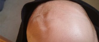
Reasons for concern
A sunken fontanel may be evidence of dehydration in a child’s body. In such cases, the help of a pediatrician is necessary. If this condition is accompanied by a serious gastrointestinal disorder, call an ambulance.
All you can do in this case before consulting a doctor or an ambulance arrives is to give your baby plenty of fluids to restore water balance.
A protruding fontanel is another good reason to see a doctor. This condition of a child for a long time may be a symptom of encephalitis or meningitis.
Anatomy lesson
The human skull is not a monolithic structure, but a collection of bones grouped into two sections: facial and brain.
The brain section, or cranium, consists of flat bones, at the junctions of which there are sutures. In newborn children, the composition and number of bones in the skull is exactly the same as in adults.
The only difference is that in an adult the brain region is twice as large as the facial region, and in a baby it is eight times larger. At the junction of several flat bones (three or more) there are holes covered with strong and dense connective tissue - a fibrous membrane.
These are fontanelles. They are named so because of the association with sources of water gushing from the depths of the earth - if you try to put your finger on a spring, you can feel the pulsation of the blood vessels.
Dimensions of a large fontanel
Most often, parents' fears are associated with the size and rate of closure of a large fontanel. Below we present the average statistical norms for its size, depending on age.
However, you must understand that this is not a dogma, and each case is individual.
The fact of deviation from the schedule does not mean anything.
- 1 month - 26-28 mm (hereinafter - width and length);
- 1-2 months — 22-25 mm;
- 2-3 months — 23-24 mm;
- 3-4 months — 20-21 mm;
- 4-5 months — 16-18 mm;
- 5-6 months - 16-18 mm;
- 6-7 months - 16-18 mm;
- 7-8 months — 14-16 mm;
- 8-9 months — 14-15 mm;
- 9-10 months — 12-14 mm;
- 10-11 months — 9-12 mm;
- 11 months — 1 year — 5-8 mm
Indeed, sometimes the fontanel behaves unusually and this may be a symptom of certain diseases.
But each of these rare diseases, which, as a rule, are genetic, has other signs that are more pronounced and immediately attract attention.
Therefore, a mother should not give her baby any complex diagnoses based only on the condition of the fontanel.
MUSCULOCAL SYSTEM
SKELETAL SYSTEM
SKULL OF A NEWBORN CHILD AND AGE FEATURES OF THE SKULL
The skull of a newborn has a number of significant features (Fig. 82, 83).
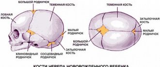
The brain skull, as a result of active growth of the brain and the early formation of sensory organs, is eight times larger in volume than the facial skull, and the eye sockets are wide. The base of the skull, compared to the vault, lags behind in growth; the bones are connected to each other through wide cartilaginous and connective tissue layers. The tubercles of the frontal and parietal bones are well defined and therefore, when viewing the skull from above, it appears quadrangular. The frontal bone consists of two halves, the brow ridges are absent, and the frontal sinus is not yet formed. The jaws are underdeveloped, which causes the low height of the facial skull. The lower jaw consists of two parts (two halves). Parts of the temporal bone are separated from each other by well-defined connective tissue or cartilaginous layers, the mastoid process is not developed. Muscle tubercles and lines are not expressed on the bones of the skull. The most characteristic feature of a newborn’s skull is the fontanelles (fonticuli), which are non-ossified connective tissue (membranous) areas of the cranial vault. There are six fontanelles in total: two are located in the roof of the skull along the midline and four are located on its lateral surfaces. The largest anterior (frontal) fontanel (fonticulus anterior, s. frontalis) is diamond-shaped, located between the two parts of the frontal bone in front and both parietal bones in the back; it heals in the second year of life. The posterior (occipital) fontanel (fonticulus posterior, s. occipitalis) is triangular in shape, located between the two parietal bones in front and the occipital scales in the back. The occipital fontanel closes in the second month after birth. The anterior lateral fontanel - the wedge-shaped fontanel (fonticulus sphenoidalis) is determined at the junction of the frontal, parietal bones, squama of the temporal bone and the greater wing of the sphenoid bone. This fontanel closes in the second or third month after birth. The posterior lateral fontanel - mastoid fontanel (fonticulus mastoideus) is located in the place where the temporal, occipital and parietal bones converge, overgrows in the second or third month of life.
Thanks to the presence of fontanelles, the newborn’s skull is elastic; its shape can change as the fetal head passes through the mother’s birth canal. It is also possible to overlap the edges of the bones of the roof of the skull one on top of the other, which leads to a reduction in the size of the skull and makes childbirth easier. This is facilitated by the Budinova plate - a cartilaginous plate located in the fetus between the lateral part of the occipital bone and its scales and causing the bones to slide and pass one after another during labor. In place of the anterior fontanel, an additional bone is often formed - Vesalius bone - the bone of the anterior fontanel (os fonticuli anterioris).
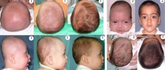
The volume of the cavity of the cerebral part of the skull of a newborn is on average 385-450 cm3. In the first 6 months. After the birth of a child, the volume of the cranial cavity doubles, by the age of two it triples, in an adult it is four times larger than the volume of the cranial cavity of a newborn. The glabella is absent in a newborn; it is formed by the age of 15. The relationship between the brain and facial parts of the skull is different in an adult and a newborn. The newborn's face is short and wide. In the lateral norm, the ratio of the areas of the facial and cerebral parts of the skull in a newborn is 1:8, in a two-year-old child - 1:6, in a five-year-old - 1:4, in a ten-year-old - 1:3, in an adult woman - 1:2.5, in adult male - 1:2.
After birth, the growth of the skull occurs unevenly, therefore, in postnatal ontogenesis, three periods of its growth and development are distinguished.
The first period - from the birth of a child to 7 years - is characterized by growth, especially in the occipital region of the skull. During the first year of life, the skull grows more or less evenly. From 1 to 3 years, the skull grows especially actively at the back. This is due to the child’s transition to upright walking in the second year of life. In the second or third year of life, due to the end of the eruption of replacement teeth and the strengthening of the function of the masticatory muscles, the growth of the facial part of the skull in height and width increases significantly. From 3 to 7 years of age, growth of the entire skull, especially its base, continues. By the age of 7, growth in the length of the base of the skull basically ends, and it reaches almost the same size as that of an adult.
The second period - from 7 years to the onset of puberty (12-13 years) - is characterized by slow but uniform growth of the skull, especially in the area of its base. At this time, the vault of the cerebral part of the skull mainly grows, especially up to 8 and at 11 - 13 years. The volume of the cavity of the cerebral part of the skull reaches 1200-1300 cm3. By the age of 13, the squamous-mastoid suture is overgrown, and the fusion of parts of the individual bones of the skull, developing from independent points of ossification, ends.
Rice. 82. Skull of a newborn, fontanelles (A - front view, B - side view, right):
1 - Mandibtilarsymphysis; 2 - Milktooth; 3 — Infra-orbital foramen; 4 - Bony nasal septum; 5 - Sphenoid; Sphenoidal bone, greaterwing; 6 - Nasal bone; 7 - Maxilla, frontal process; 8 - Frontal bone; Frontal tuber; Frontal eminence; 9 - Frontal suture; Metopic suture; 10 - Anterior fontanelle; 11 - Parietal bone; 12 - Coronal suture; 13 — Supra-orbital notch/foramen; 14 - Maxilla; 15 - Temporal bone; 16 - Zygomatic bone; 17 - Mandible; 18 — Mental foramen; 19 - Occipital bone, lateral part; 20 - Mastoid fontanelle; 21 - Lambdoid suture; 22— Squamous part of occipital bone; 23—Posterior fontanelle; 24 - Temporal bone, petrous part; 25 - Parietal bone; Parietal tuber; Parietal eminence; 26 - Sphenoidal fontanelle; 27 - Piriform aperture; 28 — Temporal bone, squamous part: 29 — Tympanio ring
Rice. 83. Skull of a newborn, fontanelles (A - top view, B - bottom view):
1 — Occipital bone, squamous part of occipital bone; 2— Lambdoid suture; 3 - Sagittal suture; 4 - Anterior fontanelle; 5 - Frontal suture; Metopic suture; 6 - Frontal bone; squamous part; 7—Corona! suture; 8— Parietal bone; Parietal tuber; Parietal cminence; 9 - Posterior fontanelle; 10 - Palatine bone, horizonta! piate; 11— Vomer; 12 - Sphenoid; Sphenoidal bone, pterygoid process; 13 - Temporal bone, petrous part; 14 - Temporal bone, squamous part; 15 - Tympanio part, tympanic ring; 16 - Mastoid fontanelle; 17 - Transverse occipital suture; 18 - Occipital bone, lateral part; 19 - Foramen magnum; 20— Choana; Posterior nasal aperture; 21 - Maxilla, palatine process; 22— Incisive bone; Premaxilla; 23 - Mandible
In the third period - from 13 to 20-23 years - the facial skull grows intensively, its gender differences appear: in boys, the facial skull grows in length more than in girls, the face lengthens. If before puberty boys and girls have a round face, then after puberty in men, as a rule, the face becomes longer, while in women it remains round.
The transformation of the skull bones in old and senile age can be attributed to the fourth period. At this time, the seams between the bones of the roof of the skull heal. The healing of the sutures of the skull begins at the age of 20-30 years, and in men somewhat earlier than in women. The sagittal suture in the posterior section begins to heal first (22-35 years), then the coronal (24-42 years), lambdoid (26-42 years), mastoid-occipital (30-81 years). The scaly seam, as a rule, does not heal. The process of healing of the sutures of the skull is individual. There are cases when in old people after 80 years all the seams were well defined.
In elderly and senile people, along with healing of the sutures, changes in the facial skull occur. Due to the wear and loss of teeth, the alveolar processes of the upper and lower jaws (alveolar arches) decrease. Due to the weakening of the chewing function, partial atrophy of the masticatory muscles, the relief of the jaws changes (smoothes), they become less massive. The bones of the skull become thinner and more fragile.
Invisible helpers
The fontanelles play a very important role from the point of view of human physiology. Firstly, they help the baby pass through the birth canal. If you look closely at the head of a newly born baby, you will notice that it is slightly flattened on the sides.
This is explained by the fact that the posterior and anterior fontanelles allowed the bones of the skull to overlap each other during the birth process and helped the little man to be born. Literally after 3-4 days the newborn’s head takes on the correct shape.
In addition, fontanelles participate in the heat exchange of the child’s body, which is very important in infancy. When the baby's temperature rises above 38°C, the membrane of the anterior fontanel helps remove excess heat from its meninges.