Causes
The main reason for violating the integrity of bone tissue is a strong mechanical impact - impact. However, the same fall can cause different injuries in children. One child will get away with a bruise, and the other will spend a good amount of time healing a fracture. The main risk factors that contribute to a decrease in bone strength include:
- Calcium deficiency. This substance is the main building material for bone. Therefore, children need to consume more foods containing calcium (milk, kefir, cottage cheese, meat, etc.).
- Sedentary lifestyle. To keep bones strong, a child must be physically active.
- Hormonal disorders. In this regard, the largest number of fractures is observed in children who have entered puberty.
Some chronic diseases can also interfere with the normal absorption of calcium. It is necessary to consult a pediatrician if your child’s hair begins to grow dull and fall out, caries develops rapidly, and the back gets tired quickly.
Injuries in children differ from those in adults. For a child, a “green stick” bone injury is typical, in which the fragments are held in place by strong periosteum that has maintained its integrity.
Damage to the joints in children is fraught with damage to the cartilage of the growth zone, and therefore the lengthening of the limb after injury may stop. In general, healing occurs faster in a child than in an adult.
This process is accompanied by the migration of cells into the pathological focus, forming a section of new bone tissue (callus) at the site of a post-traumatic hematoma. In addition, when localized in the area of the cartilaginous growth zone, necrosis of the bone tissue of the epiphysis develops in children (traumatic epiphysiolysis).
Skull fractures are most often the result of a fall from a height or a traffic accident. Both the arch and the base may be involved in the pathological process.
Some features of childhood fractures
The bones of a child contain a large amount of organic substances, in particular the protein ossein, which allows tissues to regenerate and grow together faster, unlike the bones of an adult.
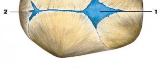
The shell covering the bone from the outside - the periosteum (periosteum) is very elastic and dense, penetrated by a large number of blood vessels, and the bone itself has growth zones - metaphyses, which have plates of cartilaginous tissue. All these nuances of the structure of a child’s tubular bones make it possible to determine the specifics of fractures.
Table. Features of childhood fractures:
| Feature of the injury | What is it characterized by? |
| According to the “green line” type | Due to the high elasticity of the periosteum, when a child is injured, the bone does not break in half, but only cracks, which looks like a broken branch of a young tree. This type of injury is characterized by a bone fracture on only one side, and on the other side the fragments are firmly held by the periosteum. As a rule, displacement of bone fragments with this type of fracture practically does not occur or they are insignificant and not dangerous |
| Bone fracture line along the growth zone | A bone fracture along the metaphysis often leads to premature ossification of cartilage tissue, which causes subsequent shortening, curvature and impaired growth of the limb. In this case, the younger the child is at the time of receiving such a fracture, the more severe the consequences will be for him and the more pronounced the shortening of the limb will be. |
| Bone outgrowth fracture | Muscles and tendons are attached to the site of the bone growth, so when such a fracture occurs, ligaments and muscle tissue are torn |
Important! The periosteum in children is supplied with blood much better than in an adult, therefore, in childhood, callus forms more quickly and the fracture site heals.
Clinical picture
The clinical picture of a fracture is characterized by the following signs:
- pain at the site of injury;
- swelling of the damaged segment;
- formation of a hematoma at the site of application of mechanical force;
Hematoma is a typical sign of a fracture in children - visible change in the shape of the limb;
- formation of a skin defect and bleeding from damaged vessels with an open fracture;
- shortening of limb length;
- loss of sensitivity and motor function of muscles due to nerve damage;
- increased body temperature;
- inability to perform habitual movements in the damaged part of the musculoskeletal system.
A skull fracture is characterized by the following symptoms:
- excruciating headache;
- episodes of loss of consciousness, convulsions;
- nausea and vomiting;
- leakage of clear cerebrospinal fluid (CSF) from the nose and ears;
- different pupil sizes (anisocoria) indicate accumulation of blood inside the cranial cavity;
Anisocoria is a sign of intracranial hematoma formation - bruising around the eyes similar to glasses.
Doctor Komarovsky about fractures in children - video
Breaks and fractures of the “green branch” or “willow twig” type
are typical injuries in childhood. With this type of fracture, observed especially often when the diaphyseal part of the bones of the forearm is damaged, the bone is slightly bent; along the convex side, the outer layers undergo a fracture, usually slit-like; along the concave side, they retain their normal structure. The bone is held in place by an intact part, primarily the periosteum.
The next characteristic form in childhood is subperiosteal fractures , which occur when force is applied along the longitudinal axis of the bone. These fractures are complete and pass in the form of a through, transverse or tortuous crack through the entire thickness of the bones. The periosteum remains intact, there is no or very slight axial displacement. The accompanying hematoma with these fractures is small. These fractures are most often observed on the bones of the forearm and lower leg.
Types of fractures depending on location
Fractures in a child are usually very different from fractures in young and old people. If the baby falls or hits himself, the following types of fractures are most often observed:
- Subperiosteal fracture;
- Epiphysiolysis;
- Osteoepiphysiolysis;
- Apophysiolysis;
- Fracture of the periosteum.
Depending on the structure and strength of the patient’s bone, the following forms of fractures are distinguished:
- Traumatic. Damage develops due to strong mechanical impact on the bone (fall, blow).
- Pathological. Such fractures can develop even with weak physical impact and are a consequence of a certain disease.
Based on the condition of the epidermis, fractures in children can be:
- closed (the integrity of the epidermis is not compromised);
- open (elements of the damaged bone violate the integrity of the skin).
Closed fractures are not infected. Open fractures have primary microbial contamination. Therefore, first aid for different types of injury differs significantly.
Depending on the type of separation of individual bone elements, fractures with and without displacement are distinguished. For young and middle-aged children, subperiosteal “green stick” fractures are most common. The peculiarity is that the damaged area does not lose the integrity of the periosteum. In most cases there is no displacement. This injury often develops in the lower leg or forearm.
Depending on the direction of the fracture line, the following types of injury are distinguished:
- star-shaped;
- transverse;
- longitudinal;
- oblique;
- helical;
- V-shaped;
- T-shaped.
Types of fractures according to location - table
According to the nature of the injury in children, fractures of the vertebral bodies are distinguished (compression, crushed, splintered), isolated fractures of the arches; transverse, spinous and articular processes of the vertebrae.
The Cunning of the Invisible Man
Loss of bone density develops slowly and gradually, it is impossible to notice it by eye. But there are five indirect signs that should alert parents.
- The child's cases of caries have become more frequent.
- “For some reason,” hair splits, nails peel and break.
- From time to time there is pain in the legs, especially in the legs.
- The schoolchild is slouching more and more, his back gets tired after a long time of sitting at homework or at the computer.
- Your child is allergic, because of this he has dietary restrictions; he does not eat dairy products or fish.
Even one such symptom is a signal that the child needs to be examined and find out whether he really does not have enough calcium.
It is a common belief that taking calcium can lead to the formation of kidney stones. This is from the realm of misconceptions. A properly selected drug is safe and also helps improve metabolism.
Causes of injuries in children
The most common injuries in young patients are injuries to the arms and legs. At the same time, about 5% are injuries to the hands and fingers. Most often, such injuries occur in children who are just beginning to take their first steps. Damage to the upper limbs can be caused by a bad fall.
Injuries in children of the first year of life are quite rare. If a child who does not yet walk or even sit is often diagnosed with fractures, it is possible that he had to deal with congenital osteoporosis.
Some babies may be diagnosed with birth injuries. Most often you have to deal with a fractured collarbone in a newborn due to the mother’s narrow pelvis. Malpresentation of the fetus is also a risk factor. Therefore, monitoring the woman and the condition of the baby in the last stages of pregnancy plays a very important role.
Children have a special bone structure. In this regard, in a child who is fully developing and does not have chronic diseases, the likelihood of getting a fracture is minimized.
Even if a young patient is diagnosed with an injury, the rehabilitation period is much faster than for an adult. Often in children of the first years of life, fractures of the bone outgrowths to which the muscles are attached are detected.
These are tears of ligaments and muscles with bone elements.
Treatment of fractures in children is complex and is carried out under the guidance of a pediatric traumatologist with the possible involvement of a neurologist and neurosurgeon. Uncomplicated cases are subject to treatment on an outpatient basis; if complications develop, hospitalization in a specialized hospital department is required.
First aid
If a child is injured, it is necessary to quickly and competently provide him with first aid. The following procedure must be followed:
- stop the action of the pathogenic factor;
- create immobilization of the fracture site using a scarf or scarf;
- in case of lack of consciousness, convulsions, vomiting, turn your head to the side;
- in case of an open fracture, apply an aseptic bandage to the damaged area;
- If your hand is injured, remove rings and other jewelry;
- in case of an open fracture accompanied by bleeding, apply a pressure bandage.
The effectiveness of first aid depends on strict adherence to the algorithm of actions
Common mistakes when providing first aid
Children rarely have bone fractures, despite frequent falls during outdoor games; however, in addition to the usual fractures observed in adults, some types of fractures appear that are characteristic only of childhood, which is explained by the peculiarities of the anatomical structure of the skeletal system and its physiological properties in children.
- The child's lower body weight and normally developed soft tissue cover weaken the impact force of a fall.
- Bones are thinner, less strong, but more elastic. Elasticity and flexibility are due to the lower content of mineral salts in the bones.
- The periosteum is thicker and richly supplied with blood, which gives the bone greater flexibility and protects it during injury.
- The epiphyses at the ends of the tubular bones are connected to the metaphyses by wide elastic germ cartilage, which weakens the force of the blow.
- Breaks and fractures like a green branch or a willow twig are caused by the flexibility of the bones.
- Subperiosteal fractures most often occur when force is applied along the longitudinal axis of the bone. The broken bone is covered by intact periosteum.
- In newborns and infants, the ossification nuclei in the epiphyses are absent or weakly expressed, so radiological diagnosis of subperiosteal fractures, epiphysiolysis and osteoepiphysiolysis without displacement is difficult. The displacement of the ossification nucleus in relation to the diaphysis of the bone can only be detected when compared with a healthy limb on radiographs in two projections. In older children, osteoepiphysiolysis is more easily diagnosed: a separation of a bone fragment is found on radiographs
- In young children, the inability to take a complete history, normally expressed subcutaneous tissue, which makes palpation difficult, and the lack of displacement of fragments in subperiosteal fractures make recognition difficult and lead to diagnostic errors
- Swelling, pain, impaired limb function, and increased body temperature resemble the clinical picture of osteomyelitis. An X-ray examination is necessary to rule out a fracture.
- A more detailed examination is often necessary, measuring the absolute and relative length of the limbs and determining the range of motion in the joints.
- The leading method of treatment is conservative: a fixing bandage is used, immobilization is carried out with a plaster splint in a functionally advantageous position, covering 2/3 of the circumference of the limb and fixing two adjacent joints. A circular plaster cast is not used for fresh fractures, as there is a danger of circulatory disorders due to increasing edema.
- Skeletal traction is often used in children older than 4-5 years.
- For displaced fractures, simultaneous closed reduction is recommended, probably earlier after the injury.
- In young children, general anesthesia should be used during reposition.
- In children under 7-8 years of age, displacement of diaphyseal fractures in width by 2/3 of the diameter is acceptable with a normal axis of the limb. In the process of growth, self-correction of such deformations occurs.
- Open reduction is performed with special care, gentle surgical access, with minimal trauma to soft tissues and bone fragments and is often completed with simple methods of osteosynthesis - Kirschner wires, extramedullary osteosynthesis.
- The time frame for consolidation of fractures in healthy children is significantly shorter.
- T14.20 Fracture in an unspecified area of the body (closed)
- T14.21 Fracture in an unspecified area of the body (open)
Typical fractures
metaphysis of the tubular bone
General principles of treatment
See also Fracture
Such fractures are a consequence of obstetric care for foot or pelvic presentation of the fetus. Typical localization is in the middle third of the diaphysis of the tubular bone; along the plane, the fracture runs in a transverse or oblique direction.
Traumatic epiphysiolysis of the proximal and distal ends of the humerus and femur are rare. This circumstance, as well as the fact that X-ray diagnostics are difficult due to the absence of ossification nuclei, often lead to untimely diagnosis of these injuries.
In diaphyseal fractures of the humerus and femur with complete displacement of bone fragments, pathological mobility at the level of the fracture, deformation, traumatic swelling and crepitus are noted. Any manipulation causes pain to the child.
Fractures of the femur are characterized by a number of features: the leg is in the typical position of flexion in the knee and hip joints for a newborn and is brought to the abdomen due to physiological hypertension of the flexor muscles.
Radiography clarifies the diagnosis. .
There are several treatment options for newborns with diaphyseal fractures of the humerus and femur.
In case of a fracture of the humerus, the limb is immobilized for a period of 10-14 days. The arm is fixed with a plaster splint from the edge of the healthy scapula to the hand in the average physiological position or with a cardboard U-shaped splint in the position of shoulder abduction to 90°.
Fracture in a child: arms, legs (feet, legs), skull, with or without displacement, how to distinguish from a bruise
Article last updated: 05/02/2018
This depends on the age and psychophysical development of the child. Preschoolers are more likely to experience household injuries, falls, and burns. Among school-age children, street and even transport injuries predominate. Riding fast on bicycles, skateboards, roller skates, and failure to comply with traffic rules leads to serious consequences.
Features of childhood injuries
Children are not a smaller copy of an adult. The structure of a child’s body, as well as the musculoskeletal system, has a number of features. Many lesions typical of children are never seen in adults, and vice versa. Why is this happening?
High percentage of soft tissue
In a growing body, the content of cartilage, fat and muscle tissue is greater than in adults. This feature has a protective function, so bones in children break much less frequently than in similar situations in adults.
High elasticity and firmness of fabrics
Due to the strength and elasticity of the periosteum, displaced fractures are rare. The periosteum, as it were, “holds” the fragments inside; such fractures are called the “green branch” or “rubber tube” type.
What is periosteum and why is it needed? The periosteum is a dense membrane that completely covers the bone. It is perfectly vascularized, supplied with blood, which means it nourishes the surface layers of the bone. Thanks to the periosteum, the bone grows in thickness.
The ligamentous apparatus of children is highly elastic. Therefore, sprains and hyperextensions of ligaments are much more common than ruptures, and dislocations before 5 years of age practically do not occur.
The special mineral composition of bones
The bones of children are thin, but they contain a lot of organic matter. Bones have great elasticity and flexibility, which also protects against fractures.
How does bone growth occur? The growth zone is a cartilaginous layer. It is located between the articular part of the bone (epiphysis) and the extension at the end of the bone (metaphysis) and allows the bone to grow in length.
Such lesions occur only in children. But it is very difficult to identify them, because cartilage tissue is not visible on an x-ray. This is serious damage that requires proper, qualified treatment and precise comparison of surfaces.
Damage to cartilage tissue
Cartilage tissue is not prone to fractures due to its homogeneous structure and elasticity. But with mechanical impact, the structure of the cartilage, its properties and content can change, and the cartilage can move and be reabsorbed.
High reducing power
Over time, a callus forms in the affected area, which is replaced by bone tissue without scar formation. And the rapid growth rate of the child allows for a “permissible displacement” to remain, which can correct itself over time.
Types of injuries in children
The most common injuries in children include bruises, dislocations, sprains, and fractures.
- Injury. How to distinguish a bruise from a fracture and other injuries? In case of a bruise, the tissue damage is minor, and its structure is not changed. Pain is the main symptom of a bruise, but it is moderate, the baby quickly calms down. Limb shape and function are not significantly changed. There might be a bruise. The bruise formed by the impact evenly permeates the tissue. The child’s condition is not significantly affected; he soon forgets about the trouble.
- Sprain. This lesion is typical for children over 3 years of age, and the typical location is the ankle ligaments. Often the baby gets this injury when running, especially on steps, when the foot turns inward. The pain of a sprained ligament is acute, but the pain gradually subsides. Edema and swelling appear in the joint area. Movement in the foot is possible, but attempts to stand on the foot are accompanied by severe pain.
- Dislocation. It often occurs when a child falls and is characterized by disruption of the normal contours of the joints. When a dislocation occurs, the ability to move the joint is severely limited. The shape of the limb changes, it becomes deformed, shortened or lengthened. Local symptoms are quite pronounced: pain, swelling, hematomas. In children aged about 2 years, subluxation of the radius in the elbow joint, “pulling dislocation,” often occurs. It occurs when an adult holds a child’s hand tightly, and the child suddenly stumbles. With such an injury, the baby cries, spares his hand, holds it along his body.
- A child has a fracture. Fracture is damage to a bone, a violation of its integrity as a result of mechanical impact.
We will talk about this type of injury in more detail.
Main causes of fractures
- falls;
- domestic injuries;
- awkward movements;
- diseases leading to disruption of bone integrity;
- mutilation.
Depending on the type of fracture, its manifestations differ, but the main symptoms are similar.
Clinical manifestations of a fracture
- pain that intensifies with movement of the limb, palpation, touch;
- deformity of the affected limb;
- unnatural position of an arm or leg, attempts to bring the limb into a physiological position lead to severe pain;
- swelling at the fracture site, which increases very quickly;
- hematoma, bruise in the affected area.
Do not forget that any injury is a damage to the entire body as a system. The body responds to damage with both local reactions (pain, hyperemia, swelling) and general ones (weakness, malaise, fever). With severe multiple fractures and injuries to internal organs, even traumatic shock may develop.
Diagnostics
How to distinguish a fracture from other types of injuries? To diagnose this type of injury, you need to know how the child was injured and under what circumstances. But parents were not always witnesses to an unpleasant situation. And a traumatized child, especially a younger one, will not be able to coherently tell the circumstances of the injuries.
It is necessary to determine the extent of damage, local reactions, abrasions, wounds, hematomas. And then pay attention to the position of the limb, whether the child can move his fingers.
Clinical manifestations of fractures can be divided into probable and reliable. Possible signs that help to suspect a fracture include swelling, hematoma, and dysfunction. Reliable ones that strongly indicate the presence of a fracture include the sensation of crunching of bone fragments and deformation of the limb.
You should always pay attention to the color of the skin around the injury, the mobility of the fingers and toes. A serious sign is paleness or cyanosis of the fingers combined with the absence of voluntary movements. This may indicate damage to a large vessel or nerve.
Another serious symptom is the absence of a pulse and unpleasant sensations in the limbs, tingling, burning, and a sensation of “pins and needles.” In such cases, it is necessary to deliver the child to a medical facility as quickly as possible.
Severe, open fractures (when the skin over the lesion is broken and a portion of the bone is visible) are rare in children. The danger of infection in such cases is great.
Even less common are gunshot, infected fractures, which require serious, long-term treatment.
More common are hand fractures of the “green twig” or “grapevine” type, which can be difficult to recognize.
An X-ray examination will help determine the exact type of damage. Only by confirming it X-ray can you be unequivocally confident in the correctness of the diagnosis. Rarely, in controversial cases, it is necessary to resort to magnetic resonance imaging.
What can be seen on an x-ray?
- the presence of a bone fracture;
- location of the fracture;
- is there a bias, is it significant;
- single fracture, or there are several fragments;
- what is the fracture line?
Child's leg fracture
- Femoral neck fracture. Sharp, unbearable pain in the hip joint, shortening of the affected limb. The leg is in an unnatural position - turned outward. And in the groin you can replace hematomas and swelling. Such symptoms indicate a displaced femoral neck fracture. If there is no displacement, the clinical picture is erased, the child can even walk.
- Patella fracture.
It is characterized by pain in the knee, swelling, and possible hemorrhage into the knee joint. The function of the leg is impaired, attempts to bend the leg cause severe pain. When the fragments diverge by more than 5 mm, the supporting function suffers and the child cannot stand on his leg. - Fracture of the leg bones.
When both bones of the leg (fibula and tibia) are fractured, deformation of the limb, severe pain, swelling and pathological mobility of the limb are noticeable. If one bone is affected, the deformity is less pronounced, and active movements in the leg are preserved. It turns out that a fracture of the tibia, depending on the number of bone fragments and their location, can be classified as either mild or severe. - Fracture of the foot bones. In addition to local manifestations of the fracture, supporting and motor functions are impaired. Movements in the foot or attempts to stand on the leg lead to sharp pain.
- Heel bone fracture. The position of the limb is changed - the heel is turned outward. Swelling and pain appear, and the inability to move in the ankle joint.
- Fractured toes.
The fingers look unnatural, are swollen, and painful when moving. There is cyanosis and hematomas under the nails. The child cannot stand on his leg.
Child's arm fracture
Fractures of the upper extremities in children are 2 times more common than those of the lower extremities. With a serious fracture, it is easy to make an unmistakable diagnosis. But for children, lesions in which the function of the hand is slightly impaired are more common. A fracture can easily be mistaken for a bruise or dislocation. The most common location is in the bones of the elbow joint and forearm.
Urgent Care
Dr. Komarovsky shares advice on providing emergency care.
What should be done:
- If there is bleeding, the first step is to stop it by applying a pressure bandage.
- Perform immobilization and fix the limb. Attach any flat object that is at hand with a bandage or cloth to the affected area.
- Apply cold.
- Immediately transport the child to the emergency room.
What not to do:
- Move the child until the limb is fixed and immobilized.
- Ask the victim to move, stand up, change seats.
- Try to change the position of the limb yourself - straighten it, align the fragments.
- Apply heat, rub, massage the damage.
Treatment of fractures
A qualified traumatologist will determine the type and amount of treatment required for each case individually. But there are basic principles for treating all fractures:
- Gentle approach, pain relief.
- Comparison of bone fragments is carried out in the shortest possible time, as quickly as possible.
- Surgical treatment if necessary.
- Fixation of the damaged limb.
- Conducting functional treatment.
Unconventional treatment and prayers for fractures are ineffective in treatment. By wasting time on traditional methods of treatment, you can miss precious minutes and harm the child. Only a qualified doctor can competently and correctly prescribe treatment.
Recovery period
How long a fracture takes to heal largely depends on the age and regenerative ability of the patient’s bone tissue.
On average, the healing time for fractures of the upper limb is one to one and a half months, and for fractures of the lower limb - 1.5 - 2 months.
Healing of a fractured pelvic bone will require even more - from 2 to 3 months, and for the spine the healing period will last up to 1 year, and complete recovery - 2 years.
Healing time also depends on the type of fracture and treatment tactics. For example, with a simple fracture of the leg bones, a plaster splint may be applied for a period of 6 to 7 weeks. But in cases where it is impossible to combine the fragments with your hands, they resort to reposition using skeletal traction for a period of 4 - 8 weeks, followed by plaster casting. This means that the healing time of the shin bones doubles.
After removing the plaster cast, the active recovery period begins. The best methods of therapy at this time are massage, physical therapy, physiotherapy, and a swimming pool.
Do not forget about proper nutrition of the child and the increased need for microelements during the recovery period. Vitamin-mineral complexes, which include calcium, will increase regenerative processes and accelerate fracture healing.
In case of severe injuries, sanatorium treatment and long-term rehabilitation may be required.
conclusions
All children are injured. This is a payment for the curiosity and activity of the baby. No matter how loving and caring parents are, they are not able to protect the baby from all possible troubles.
The parents’ task is to recognize the injury, be able to provide emergency assistance and, if necessary, transport it to a medical facility in a timely manner. The health and development of the child in the future depends on the correctness and speed of providing first aid and medical assistance.
Source: https://kroha.info/health/disease/perelomy-u-detej
How to recognize a fracture
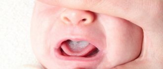
It is not difficult to suspect a fracture in a child. Immediately after the injury, the baby feels sharp pain and cries. The site of injury rapidly swells and acquires a bluish tint. A characteristic sign of a limb fracture is its deformation. In addition, the child may become pale, sticky sweat appears, and body temperature rises to low-grade levels.
Nonspecific symptoms may occur with greenstick fractures. The child can maintain physical activity and there will be virtually no pain. Often, it is possible to determine the presence of a fracture only with the help of hardware diagnostics in a hospital.
Treatment
If a fracture is detected in a child, it is strictly forbidden to self-medicate. Therapy should only be prescribed by a qualified specialist. At the same time, parents should know how to provide assistance to the victim before the ambulance arrives. The algorithm of actions should be as follows:
- Immobilize the damaged area using a splint. Any hard means at hand will do - a ruler, a board, a stick. As a last resort, you can roll up a magazine. If the tire turns out to be quite rough, wrap it in a bandage or towel before applying it. If a rib is fractured, a pressure bandage is applied.
- It is necessary to ensure that the splint is applied above and below the fracture joints.
- The splint should be carefully secured using a bandage. The bandage should not be too tight.
- To relieve pain, the child can be given a drug based on ibuprofen or paracetamol.
In case of an open fracture, before immobilizing the damaged area, it is necessary to treat it with an antiseptic and stop the bleeding. It is advisable to carefully remove clothing from the area of injury (it is better to cut it off).
In case of an open fracture, the doctor must clarify whether the patient has previously been vaccinated against tetanus.
For simple fractures without displacement, the prognosis of therapy is usually favorable. Children's bones heal quickly and their function is restored. The rehabilitation period in most cases does not exceed 3 months.
Displaced bone injuries require longer rehabilitation. It is often necessary to perform multiple surgeries to restore normal functionality to the injured area. The following complications are possible:
- injuries to nerves, ligaments and tendons;
- addition of a bacterial infection;
- improper fusion of bone, which leads to disruption of its functionality.
In most cases, if qualified assistance is provided in a timely manner, the child’s health condition is completely restored. However, unpleasant consequences of fractures are also possible. The most common complication is premature closure of the growth plate, resulting in deformed bone.
It is not always possible to protect a child from falls and injuries. But you can significantly reduce the likelihood of fractures if you monitor your baby’s diet. Food should be healthy and varied.
Physical activity is also of great importance. The child should regularly spend time in the fresh air and experience moderate physical activity.
How to help a child with a fracture?
Child's arm fracture
If you suspect a fracture in a child, then it is necessary to take the victim to the hospital - the doctor will assess the severity of the injury and prescribe adequate treatment with minimal consequences for musculoskeletal function in the future.
Principles of providing first aid for a fracture
The main aspect of providing assistance to a victim with a fractured limb is to ensure its immobility so that the bone fragments do not move and injure healthy areas of tissue.
Below are instructions for providing first aid to patients with a closed fracture:
- Give the victim an anesthetic - since a fracture is accompanied by severe pain, before proceeding with fixation of the limb, it is necessary to reduce the pain threshold in order to avoid the development of pain shock. For a child, preparations based on Nimesulil or Ibuprofen are ideal - Nurofen, Nimesil, Nemidar and others.
- Securely fix the injured limb - this prevents bone displacement and injury from soft tissue fragments. If you don’t have any special devices at hand, you can fix the broken bone with two flat boards, wide rulers or plywood, tightly bandaging it. Important! Not only the broken bone should be fixed and immobilized, but also the adjacent joints, for example, in the case of a forearm fracture, the wrist and elbow.
- Take the victim to the hospital.
First aid for open fractures
Ambulance
A fracture with a violation of the integrity of soft tissues requires special attention and caution in actions, since protrusion of the broken bone outward is accompanied by bleeding and a high risk of further complications.
If the wound is large and the bleeding is massive, you should quickly navigate and determine its type - venous or arterial, on which further actions will depend. With venous bleeding, the blood, which is a dark, rich red color, usually flows calmly. With arterial blood, the blood is scarlet, bright, gushes and quickly leads to life-threatening complications.
Important! In case of an open fracture in a child with bleeding, first of all, you should give an anesthetic and only then quickly proceed to other manipulations.
To stop venous bleeding, a tourniquet or tight bandage is applied to the victim below the fracture site, always including a note indicating what time and by whom these actions were carried out. In case of arterial bleeding, a bandage is applied above the fracture site with the same note, after which they begin to treat the wound surface and fix the limb.
Important! If there is no bandaging material or rope suitable for the role of a tourniquet at hand, then you can tear the clothes into strips or firmly press the place of the torn vessel with your fingers and wait for an ambulance - all actions must be quick, since the price of delay or panic is the life of the child.
Open fractures pose a threat of infection, so a mandatory condition for providing first aid to the victim is the administration of anti-tetanus serum. Treatment of fractures in children is usually carried out on an outpatient basis; hospitalization is required only if surgical reposition of the fragments is necessary.
Treatment of fractures in children
https://youtu.be/w5vEbaOL3bQ
Complications
The prognosis for treatment of fractures in children largely depends on the nature of the injury. Multiple injuries, crushed bones with complete loss of some areas complicate the situation. In severe cases, the following complications may develop:
- systemic reaction of the body to damage - traumatic shock;
- the body's systemic reaction to blood loss is hemorrhagic shock;
- wound suppuration;
- post-traumatic purulent inflammation of the bone - osteomyelitis;
- non-union of the fracture with the formation of pathological mobility of the bone area (false joint);
- formation of joint stiffness;
- limb deformity;
- shortening of the limb with the formation of lameness;
- death.
Prevention
It is necessary to explain to children the rules of safe behavior on the street, at home in child care institutions, and in transport. Young children require supervision.
There should be no dangerous objects that could cause injury in the room where the child is located. Small children must be transported in a car using a special restraint device.
Parents should feed their baby foods rich in calcium and phosphorus, as well as foods high in vitamins and minerals.
Norm and deviations
First of all, the pediatrician will prescribe biochemical tests of blood and urine, which can be used to determine whether phosphorus-calcium metabolism is impaired. These partner minerals are involved in many vital metabolic processes and work hand in hand: the body cannot absorb calcium if there is not enough phosphorus, but if there is an excess of the latter, calcium is excreted from the body. That's why it's so important to maintain their balance. By comparing the data with the standard indicators for a certain age and detecting deviations, one can suspect the initial stage of osteopenia.
To clarify the diagnosis, densitometry is performed: assessment of bone tissue (ultrasound is often used). Unlike adults, children are analyzed only by the so-called Z-criterion - that is, deviations from the norm in indicators depending on the age and gender of the young patient, which are calculated using a special computer program.