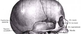
The skull of a newborn baby is different from that of an adult. The skeleton bones of the baby's head have soft spaces (sutures) and membranous non-ossified zones (fontanelles). These soft areas allow the baby's head to pass through narrow spaces in the birth canal. As a result of labor, cranial deformation occurs, which is observed in most newborns.
After birth, the baby takes his first breath in a process that expands not only the lungs, but also the cranial part of the skeleton. Then, when sucking, the wedge-shaped occipital joint acts as a kind of lever, creating pressure that can even out the asymmetry. However, there are congenital and acquired deformities that require treatment.
Head shapes
The baby's skull may be injured during childbirth
In the first months of life, a baby's head may appear too large or small, uneven or asymmetrical. Up to 6 months, the process of active growth and development affects the shape of the skull. Normally, the head circumference reaches 32-38 cm. In children under 6 months, every 4 weeks the parameter increases by 1.5-2 cm, and then the growth rate decreases to 5-10 mm.
Indicators of normal form may also differ. In pediatrics, there are several options for skull deformation in newborns:
- Brachycephaly. There is a flat forehead and widening of the bones of the crown, the occipital part protrudes forward. Common causes of the disorder include infections and trauma during childbirth.
- Acromegaly. The deformity is accompanied by a strong increase in the skull on both sides. Among the reasons are hormonal diseases due to cancer in the pituitary gland. A large nose is often observed.
- Scaphocephaly. Deformity with displacement of the frontal or occipital bone forward. Sometimes accompanied by impaired growth and development of the brain.
- Dolichocephaly. Deformation with a narrow and elongated head, “egg-shaped” crown, high forehead. Most often occurs after a rapid process of delivery. Usually it returns to normal on its own.
- Plagiocephaly. When deformed, flat side surfaces remain. The size of the skull is larger than normal, as is the weight of the fetus itself.
- Microcephaly. The head is too small, which is why brain development is quickly impaired. Often bones fuse prematurely and developmental delay occurs.
- Trigonocephaly. The deformity affects the frontal part. It becomes pointed, and the lower jaw moves forward and increases in size. It is formed due to premature fusion of the cartilage of the skull.
- Hydrocephalus. A pathology in which there is high cranial pressure and the head itself is enlarged in size. Just like microcephaly, it has dangerous consequences.
The key factor that provokes head asymmetry in a child is the characteristics of the birth process. In most cases, after a cesarean section, the head acquires a brachiocephalic shape. This is one of the indicators of the norm, which after childbirth will require a small home correction. During natural childbirth, in the first days, the newborn’s head has a dolichocephalic shape.
In the first months of life, the baby’s skull bones are characterized by softness, and their shape gradually changes. Normally it becomes round. However, pathological processes can interfere with the natural formation of contours.
What factors provoke pathologies?
Minor irregularities in the skull may resolve on their own within 2 to 3 months.
Small irregularities and deformations are straightened out when the child is 2-3 months old and begins to sit up. However, some deviations can accelerate bone fusion, which often causes the development of pathologies. The reasons for these processes include:
- bone dysplasia caused by genetic changes;
- hereditary deviations;
- abnormal position of the fetus during pregnancy;
- intrauterine infections.
Less common factors leading to skull deformation in a newborn include:
- combination of a large fetus with a narrow birth canal;
- atony of muscle tissue in a woman in labor;
- narrow or flat pelvis;
- 2 or 3 fruits;
- expulsion of the fetus too quickly;
- incorrect insertion of the head into the pelvic ring.
There is also a group of causes of acquired asymmetry, which occurs when the child is incorrectly positioned in the crib in the first weeks of life. Acquired deformity can occur when the baby is not latched to the breast correctly and is carried incorrectly in the arms.
If a baby has a sloping head that is stretched upward, this in 99% of cases indicates an incorrect position in the crib. A flat back of the head can be a sign of rickets. Sometimes the bevel is observed to the left or to the right. This occurs due to the child remaining on one side for a long time.
How to straighten a newborn's head?
Deformation of the bones of the baby’s skull at an early stage, until the final closure of the fontanelles has occurred, can be corrected using craniosacral therapy. The doctor uses manual techniques to eliminate the asymmetry of the baby’s skull. Massage for head deformation of a child under 1 year of age has shown the effectiveness and safety of therapy, provided the doctor is highly qualified.
Parents can eliminate the anomaly on their own:
- avoid incorrect positioning of the baby during feeding;
- periodically change his position in the crib, placing him on the other side, on his tummy;
- move the crib relative to the light and sound source.
A newborn with congenital asymmetry of the skull should be periodically examined by a pediatrician or neurologist. A small child responds more easily to conservative treatment methods. If correction by such methods is ineffective, then the doctor selects an orthopedic correction helmet for the baby. It determines the size, frequency and duration of wearing the device.
Correction of a child's head using an orthopedic helmet
Parents should remember that strict adherence to the specialist’s recommendations, perseverance and consistency in performing procedures will help completely correct the injury, the child’s skeleton and avoid complications that can result from asymmetry of the baby’s head. In particularly severe cases, surgery may be required.
Signs of a deformity causing problems
Persistent deformities require medical or surgical treatment
A visually uneven skull in a child aged 3-6 months does not always make one think that this is a pathology. And even minor deformations can cause compression of blood vessels, nerve endings and brain tissue. They can be suspected by the following symptoms:
- strabismus develops;
- the child spits up during and after meals;
- during feeding, an incorrect sucking process is observed;
- torticollis develops;
- intracranial pressure increases;
- movement coordination is impaired;
- hearing problems appear;
- lethargy and muscle weakness are observed.
The most dangerous symptoms are: jaundice, soft and hard bumps on the skull, purple or bluish spots, lacrimation, asymmetry of facial expressions and jaws, bulging of the fontanel in a calm state. These signs require immediate consultation with a doctor and instrumental diagnostics.
Symptoms
Head deformation is a serious pathology that causes various complications. Usually the diseases are accompanied by neurological disorders. Deformation causes physiological, psychological and anatomical disorders.
May be noted:
- headache;
- decreased memory and attention;
- loss of consciousness;
- attacks of irritability and sudden aggression;
- cramps and spasms of facial muscles;
- nystagmus;
- vomit;
- a sharp change in emotional background;
- strabismus;
- exophthalmos;
- torticollis;
- curvature in the area of the eye sockets, cheekbones and jaw;
- lack of hair growth in the affected area;
- asymmetrical arrangement of the ears;
- malocclusion;
- growth retardation.
Pathology significantly reduces the quality of life of an infant. Therefore, timely diagnosis is important. Based on the data obtained, the specialist draws up a suitable treatment regimen.
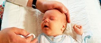
Examination by a neurologist and examination
During the examination, the doctor measures the circumference and sagittal size of the head.
During a routine visit to the pediatrician, pathological asymmetry can be detected in 99% of cases. Next, a referral to a neurologist or neurologist is written out. In difficult cases, consultation with a neurosurgeon is required. If you suspect any abnormalities within 1 month, you can immediately contact a neurologist. It is recommended to do this even in the absence of symptoms of the disorder - to exclude hidden pathologies.
If a newborn’s head deformity is confirmed, the doctor interviews the parents and prescribes the following procedures:
- X-ray. With its help, features of deformation are identified. The technique is used if there is no doubt about the presence of a pathological bone structure.
- MRI or CT. Used in cases where the diagnosis is unclear or there are doubts about its reliability.
- Vascular Doppler. Detects compression of veins and arteries.
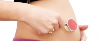
- Ultrasound. Used to determine deformities during pregnancy.
Only after studying the test results can the doctor prescribe a suitable method of therapy.
Diagnostic methods
MRIs are performed on infants under light anesthesia.
A diagnostic examination involves an initial visual examination of the patient and a detailed study of the medical history. In order to determine anatomical disorders, a specialist palpates the skull.
Also used:
- venography;
- Ultrasound (ultrasound examination) of internal organs;
- angiography of cerebral capillaries;
- CT (computed tomography);
- MRI (magnetic resonance imaging).
During the examination, the duration of development of the disease is taken into account. Genetic diseases in the family are identified.
Treatment options
Treatment of irregularities with orthoses
Depending on the type of head deformation in the child, recovery tactics are selected. These may be medications in the case of dropsy, surgical interventions for dangerous disorders, and home methods for non-pathological curvature of the bones.
Use of medications
Drugs are prescribed for hormonal disorders, especially changes in the functioning of the pituitary gland. They may also be needed for immune disorders, as well as during the recovery process after surgery.
Cranial orthoses
Special orthopedic products aimed at restoring a crooked head in a child. Selected individually. Their direct purpose is improper bone formation and asymmetry. The orthosis creates constant but slight pressure in the desired part of the head. Orthopedic devices are prescribed from 4 to 6 months of life, when the bones are still soft, but do not grow as quickly as in the first 12 weeks.
They wear such a bandage constantly, and take it off only for a while - no more than once a week.
You can use orthoses even at an older age. But in this case, their shape and period of wearing will change. If in the first year of life they are prescribed for a maximum of 4 months, then in adulthood it takes up to a year to recover.
Tips for parents with minor asymmetry
During sleep, the baby's head must be turned periodically
If the doctor does not find any pathological abnormalities, he may prescribe home treatment. It comes down to simple everyday procedures and rules:
- carry the child in your arms more often if he is not sleeping, to reduce pressure on the bones of the skull;
- when resting, change the position of the newborn’s head, tilting it to the left or to the right;
- put the baby on his stomach more often, observing safety precautions and not leaving him unattended;
- sign up for a relaxing and strengthening massage course with a professional;
- put the child in different positions more often so that he independently changes the position of his head;
- Apply alternately to the left and then to the right breast when feeding.
It is useful to have a special orthopedic pillow for newborns in your arsenal.
Performing an independent massage, especially in the head area with deformation, is strictly prohibited. Even a little incorrect pressure can make the situation worse.
Working with an osteopath
If parents turn to an expert in the first year of a child’s life, the chances of recovery increase tenfold. An osteopath helps eliminate hypertension, relieves spasms and helps the proper development of the cervical spine. If it functions correctly, all organs receive sufficient nutrition and oxygen.
Surgical intervention
Premature fusion of the skull bones is corrected surgically
The indication for surgery is premature fusion of the skull bones. This is required in approximately 7% of all head deformity cases. The procedure is simple and carried out quickly. Children recover within 2-4 months. Also prescribed when there is a risk of brain compression.
To distinguish pathological deformation of the skull in a child from natural processes that do not require medical intervention, it is important to undergo professional diagnostics. If violations are detected, the doctor will be able to quickly select effective treatment. After it, children do not experience developmental problems.