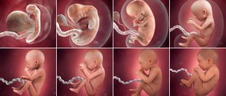
The most wonderful event for a woman is the first meeting with her child, whom she carried within her for 9 months and all this time she only guessed what he would look like. But finally the moment of childbirth comes, and the long-awaited meeting takes place. Probably every mother carefully studies the appearance of her child, and if she pays attention to other babies, she will notice that not everyone has the same skull shape. In this regard, the question may arise: why?
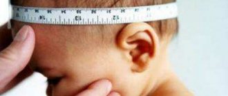
Baby's head circumference
In the first 12 months of his life, the baby grows and develops rapidly. The process also applies to the baby’s head - during this period the diameter of his skull should increase by several centimeters!
The greatest activity of this stage is observed in the first half of life. In a one-month-old baby, compared to a newborn, the diameter of the skull will increase by 2 cm! This process slows down only by the 4th month of life.
Parents sometimes think that the fetus has a big head. There is nothing pathogenic or scary about this. The baby's body will acquire the correct proportions only by one year. But at the 15-16th week of life, his chest and head will be completely the same diameter.
What is the normal skull circumference for a newborn?
The normal head circumference of a newborn baby is 35 cm. However, taking into account individual characteristics, the normal diameter of the skull will be in the range of 32-38 cm. Further observation is carried out taking into account the size of the head circumference at birth.
If the indicators are slightly higher than normal, then, accordingly, during subsequent development, a small increase will be normal. If the head size at birth is smaller than standard, then this should be taken into account when analyzing development indicators.
Microcephaly
The second type of possible pathology in newborns is a disease such as microcephaly. With it, there is a decrease in brain mass in a newborn, in contrast to healthy children, and an associated decrease in the size of the head circumference.
There are many reasons that provoke the development of this disease. These can be various infectious diseases suffered during pregnancy, intoxication of the fetus in the womb with alcohol, tobacco and drugs. Such effects are especially dangerous in the early stages of pregnancy, when all the organs and systems of the child are just being formed.
The use of certain antibiotics during pregnancy has a negative effect. The influence of radioactive radiation, toxic poisoning of the fetus, genetic abnormalities and birth injuries can also cause the development of microcephaly in newborns. In this case, the child’s skull will be noticeably smaller compared to children without pathology.
Table with baby head sizes
The table specially compiled by scientists “Child’s head circumference by month” is very helpful here. You can read it in the article. The table not only shows the norm for a certain age, but deviations from it that are not pathological.
However, the table “Child’s head circumference by month” is not able to reflect the individual characteristics of each baby. Therefore, measurements should only be taken by a pediatrician, analyzing the results regarding a specific baby.
Each parent can independently calculate the normal increase in the size of their child’s skull as they grow and develop:
- Children 0-6 months have the fastest increase in head diameter. Every month it normally increases by 1.5-2 cm.
- It is considered normal for babies 0.5-1 years old to increase their head circumference by 0.5-1 cm every month.
Pathological and non-dangerous deviations in head size
Why is it so important to monitor the increase in the diameter of the baby's head? Sizes that are too small or too large may indicate a serious illness. But deviations are not always pathological in nature. The main reasons are as follows:
- Hydrocephalus. This is a pathologically large head in a newborn. A congenital defect that causes dropsy of the brain. It leads to swelling of the baby’s fontanel, an increase in the size of the skull, and a characteristic protrusion of the venous network on the head. The danger of the defect is that it can lead to both serious neurological disorders and death.
- Microcephaly. With this pathology, the baby, on the contrary, has a very small head. A closed fontanelle does not allow the skull to expand. Such developmental delay is fraught with a wide range of consequences.
- Consequences of birth trauma. One of the common reasons. When passing through the birth canal, the child could touch not only the internal tissues of the mother, but also hit the bones with his head through their thickness. The injury leads to the appearance of edema. In most cases, this effect goes away on its own within 24 hours. But a certain percentage of children require serious treatment. Swelling at some point makes the child's head larger than normal.
- Hereditary factor. If most of your family members have a somewhat large or small head, then it is not surprising that the newborn heir will boast of a similar feature. You should warn your pediatrician about this fact.
Symptoms of microcephaly
Microcephaly of a newborn can be recognized even visually, without additional examinations. This disease is accompanied by the following symptoms:
- The head circumference of a newborn is 2-3 times smaller than the norm. If in healthy children it is 32-38 cm, then in newborns with microcephaly this figure is only 25-27 centimeters. Photos of newborn children with microcephaly show that the shape of their skull is changed - the child’s face grows, but the head itself remains small.
- The weight of the brain in healthy children is approximately 400 g, and in newborns with microcephaly it fluctuates around 250 g.
- Frequent companions of this disease are such deviations as “cleft lip”, strabismus, “cleft palate”.
- Children with microcephaly are born with a closed fontanel, or its closure occurs in the first month of life.
- The baby is noticeably lagging behind in emotional and speech development. At the same time, he not only cannot reproduce words and sounds himself, but also practically does not understand the speech spoken by others.
Microcephaly is currently, unfortunately, an incurable disease. Treatment is mainly aimed at reducing the development of defects.
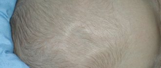
An uneven head is normal!
Let's move on from sizes to skull deformations in children. Experts tell us: an uneven head in a baby is absolutely normal!
The fact is that the fetus’s body, like its mother, is also preparing for the upcoming birth. That is why, until birth, wise nature leaves the bones of the child’s skull soft. This helps him pass through the narrow birth canal more easily.
If a woman gives birth to a baby naturally, his head is normally slightly deformed or enlarged. Children born by caesarean section, as a rule, will not have this feature.
The flat head of infants at birth is somewhat elongated, which leads to deformations of the skull in children and the appearance of small irregularities. There is no need to worry here. In the process of development and growth, the asymmetry will pass, and the irregularities will smooth out.
A child’s head becomes round and even in shape only by one year of age. And the final circumference of the skull in most children is formed only by school age.
Why was the child’s skull deformed?
Skull deformation in infants can occur not only at birth. Sometimes parents notice that during development, the child’s skull has changed unnaturally. What's happened?
Let's look at the most common cases:
- Strongly elongated or sloping back of the head. In this case, the head may be uneven, flattened, and its size no longer corresponds to normal. What does this type of skull shape indicate? The problem most often is that the baby is in the same type of lying position for too long. Newborns have the peculiarity of tilting their head to a certain side. This leads to the development of skull deformation in children.
- The baby’s skull bones remain soft for a long time. This is provided by nature for a reason: this feature allows the brain to develop unhindered and protects the child himself from injury. Therefore, if he often turns his head in a certain direction, lies on one side, all this can affect the shape of his skull. Mothers always move the child from one position to another, placing him in different directions from the object of interest.
- Don't forget about the fontanel. This is an area on the head characterized by elastic soft tissues. While the fontanel is open and not closed, the shape of the child’s skull can sometimes change significantly. The head becomes lopsided or flat if the baby just lies in the same position for a long time. Therefore, parents should always pay attention to this fact so that in the future the adult child does not blame them for his disproportionate skull shape.
A child has a crooked head
For 10 years, I have been successfully treating children with perinatal damage to the central nervous system, and they often ask me to straighten a child’s crooked head. They go not only to me with this problem, but also to other doctors - osteopaths, craniosacral therapists... they go to various grandmothers... grandfathers... and they correct and adjust the child’s head, but it just doesn’t straighten out... and they are looking for someone who is still... will cure and heal this illness...
The desire of parents to help their child is quite understandable and understandable, but this is not what we are talking about. Since this topic has long been overgrown with myths and speculation, I want to clarify some points on this topic, and therefore we will talk about the reasons for such deformations, and whether it is really possible to help in this situation. I think that this will be useful both for parents of children with this problem, and for specialists who work with this pathology.
Before we get to the causes of deformation, I will dispel one myth!
Among the people, and even among doctors, there is such a concept - “the child has laid his head to rest.” He was lying in the crib in such a way that he was always looking in one direction, or the child has torticollis and the head is turned to one side, and therefore the back of the head is flat, but in a healthy baby this will not make the back of the head flat - this is a myth!
For this to happen, you need to lie motionless with your head turned to the side for a long period of time, without changing the position of your body and head, and the child does not lie in one position, he is picked up, swaddled, turned over on his stomach, bathed, fed etc., and, in fact, with a regular change of position, the head cannot be deformed, it simply does not have time to do this, but, nevertheless, there is a nuance - if the child has paresis of the neck muscles, decreased tone in the arms (as a result of hypoxia and ischemia of the cervical thickening and medulla oblongata), vitamin D deficiency (Rachitis is a disorder of bone formation and softening of bones), and the child lies on his back every day, throughout the day, then the skull may become deformed, and the back of the head will become flat, which will indicate perinatal damage to the nervous system and vitamin D deficiency.
A child without perinatal damage, without hypoxia and without vitamin D deficiency will not have skull deformation, even if he often lies in one position.
Of course, if desired, you can give a certain shape to the skull; to do this, you need to rigidly fix the child’s head for a long period of time (artificial deformation of the skull) using tight bandages-caps or special structural wooden devices, this is what various tribes of South America used to do - Paracas, Nazca, The Incas; North America; Congo; Sudan, who deliberately deformed their heads in order to become like the gods. Nowadays, there is a similar method for correcting the shape of a child’s head using special orthopedic helmets, but they have not justified their effectiveness.
Now about the causes of deformation, there are two of them:
- Congenital developmental anomalies (craniostenosis).
- Tension (strain) of the dura mater.
Craniostenosis (congenital developmental anomalies) is premature, intrauterine ossification, fusion of the sutures of the skull.
Depending on which suture fuses prematurely, a certain deformation and shape of the skull occurs.
Normally, in a newborn up to 2 years of age, the cranial vault is mobile, sutures are formed by 5–7 years.
A child is already born with a certain deformation of the head, and as the child grows, during the first months, the deformation becomes even more pronounced.
The presence of congenital developmental anomalies in a child indicates hypoxic, infectious or toxic damaging effects in the first five months of intrauterine development.
Types of craniostenosis:
- Scaphocephaly is fusion of the sagittal suture (Fig. 1). The skull is narrowed and elongated, with a large longitudinal and small transverse diameter of the brain skull.
- Trigonocephaly is a fusion of the metopic suture (Fig. 2). Triangular or keel-shaped head.
- Plagiocephaly is the fusion of the lamboid suture (occipital variant) or coronal suture (frontal variant) on one side.
In the frontal variant, there is a flattened frontal bone on the right or left and an oblique shape of the skull (Fig. 3).
With the occipital one-sided variant, there will be a beveled, flat occiput on the right or left and an oblique shape of the skull (Fig. 4).
- Brachycephaly is the fusion of two coronal or two lambdoid sutures at the same time (Fig. 5).
Short head, large transverse and small longitudinal diameter of the cerebral part of the skull.
- Oxycephaly is ossification of the sutures between the frontal, parietal, occipital and temporal bones (Fig. 6), with compensatory growth of the bones of the calvarium in the area of the anterior fontanelle. Pointed, conical shape of the skull.
There is another form of the skull - Dolichocephaly (Fig. 7), but this is not a developmental anomaly, there is no ossification of the sutures (a normal variant). The shape of the skull is elongated.
With craniostenosis, no matter how you try to straighten the child’s head using craniosacral therapy, osteopathy, biodynamics and other conservative treatment methods (massage, manual therapy), unfortunately, the deformity will not disappear, and the healer’s hereditary grandmother will not help here either.
As a rule, deformation of the skull does not affect the mental and physical development of the child in any way and does not require special treatment, but since it can still manifest itself as an externally aesthetic defect on the face in the form of asymmetry of the facial skull, it is possible to use surgical treatment of craniostenosis to eliminate this defect.
Oxycephaly requires surgical treatment, otherwise both mental and physical development will suffer, since prematurely fused sutures of the skull will not allow the brain to increase in volume as the child grows.
Tension of the dura spinal membrane
The dural spinal membrane (Fig. 1.1) is a sac consisting of collagen tissue surrounding the brain, spinal cord, as well as spinal nerves, each of which is formed by the anterior (motor) and posterior (sensitive) roots of the spinal cord, which at the exit through the intervertebral foramina are connected, and a spinal nerve with sensory and motor fibers is formed (Fig. 2.1).
The spinal membrane covers and accompanies each nerve and passes into the nerve sheath, and any tension and tension from the internal organs will be transmitted to the spinal membrane and cause strain or torsion (Fig. 3.1), depending on the level of localization of the primary fluidic tension (FN) in the zones of the ventral axis (in the chakras). FN has a centripetal effect, attracts similar quality fluids and water and creates secondary tension of the spinal membrane.
Fixation and accumulation of fluid at the level of autonomic ganglia, plexuses is manifested by edema and the formation of persistent dominants, at the level of arteries it is manifested by spasm in diameter and along the length, at the level of hollow visceral organs (small, large intestine, stomach, excretory ducts, sphincters) it is manifested by spasm and disturbance peristalsis in the ducts and intestines.
Since the dura mater, namely the falx and tentorium of the cerebellum, is connected to the vault and base of the skull, the tension of the spinal mater causes secondary deformations of the bones of the skull and deformation of the head occurs.
Such deformities are successfully corrected using fluidic and osteopathic techniques (on average, 2–4 sessions are needed), and the child’s head gradually straightens and acquires a normal shape.
Tension of the dura mater comes from the ventral axis, along the spinal nerves.
The ventral axis is located anterior to the anterior surface of the vertebral bodies and is conventionally divided into zones.
These zones correspond to the chakras or diaphragms or rachian belts.
Tensions in the zones arise secondary, due to negative feelings, generic tendencies, dead energies, stressful situations, ego and psycho-traumas, unresolved life situations (present and past lives), gestalts, planetary influences, distorted, interrupted or rigid flows .
Anatomy of zones (chakras) (Fig. 4.1.1 and 4.2.1)
- Zone 1 (Ajna) - includes the medial part of the orbits, the ethmoid bone with a perpendicular plate, nasal bones, vomer, cockscomb, anterior part of the falx, olfactory receptors, olfactory bulbs, mediobasal sections of the frontal lobes; pyramids of the temporal bones, base of the wings of the sphenoid bone (Fig. 5.1, 5.2, 5.3).
- Zone 2 (Vishuddha) - includes the lower jaw, floor of the mouth, esophagus, trachea, aortic arch, brachiocephalic trunk, common carotid arteries, subclavian arteries, autonomic plexuses, lungs (Fig. 6.1, 6.2, 6.3)
- Zone 3 (Anahata) - includes the posterior mediastinum, thoracic diaphragm, thoracic aorta, thoracic lymphatic duct, renal arteries, celiac trunk, superior mesenteric artery, pancreas, liver, spleen, autonomic ganglia, thoracic plexuses, kidneys (Figure 7.1 , 7.2);
- Zone 4 (Manipura) - includes the mesentery of the small intestine, small intestine, inferior mesenteric artery, metasympathetic nervous system of the abdomen, sigmoid colon, cecum, inguinal ligaments, pelvic nerve plexuses, ovaries (in women) (Figure 8.1).
- Zone 5 (Svadhisthana) - includes the urogenital diaphragm, symphysis, bladder, prostate (in men), uterus (in women), ampulla of the rectum, arteries, veins, nerves and bones of the lower extremities (Fig. 9.1, 9.2).
Generic tendencies (Fig. 10.1), psycho-emotional overload, negative feelings, gestalts lead to secondary tension in the zones (chakras) and, as a consequence, tension in the dura mater, which can occur even in the perinatal period (intrauterine), which is predisposing factor in the occurrence of intrauterine hypoxia in the perinatal period, birth trauma in the natal period and chronic hypoxic process in the postnatal period.
Therefore, it is advisable to carry out timely psycho-somatic correction very usefully and effectively:
- in the perinatal period (1st, 2nd and 3rd trimesters of pregnancy) for both the woman and the child, thereby greatly reducing or preventing the likelihood of developing birth injuries (hypoxic and mechanical),
- and at an early stage of the baby’s development (from birth to one year), the sooner psychosomatic and osteopathic correction is carried out, the faster the baby will recover from injuries and develop in accordance with age.
In the first year of life, the child’s nervous system is still maturing, myelination of nerve fibers occurs, and this is the most favorable and effective period for correcting perinatal problems of the child’s nervous system!
Don't miss this opportunity, let your child be healthy!
When does the danger of deformation pass?
Don’t think that you will always have to monitor how often your baby turns his head to the right or left, or which side he lies on more often. Pediatricians reassure: deformation of the skull in an infant is a phenomenon characteristic of the period when he can only lie down.
As soon as the baby learns to sit down and begins to spend more time in an upright position, the situation will change. As a rule, already in the 2-3rd month of life, the baby’s skull becomes noticeably straighter, deformations disappear, and the head gradually begins to take on a permanent correct shape.
By the way, the opposite problem here will be the overgrowth of fontanelles too quickly. In this case, the skull becomes prematurely rigid. Of course, this saves the baby from the danger of skull deformation earlier, but it is fraught with other things. The child suffers from increased intracranial pressure.
Rickets
Rickets is a disease that still occurs quite often in young children. Deformation of the head shape is one of its most common manifestations.
The cause of rickets is a lack of calcium in the body. Because of this, the baby grows and develops slowly, his bones are weak and fragile. Another consequence is that the fontanelles do not heal for a long time. Therefore, the cranial bones remain soft even in a relatively large child. And, as a result, they remain susceptible to deformation longer.
Treatment is prescribed in the form of taking medications containing calcium and vitamin D. It is also important to introduce foods rich in these elements into the baby’s diet and spend time with him in the fresh air more often.
Causes of rickets in children
Rickets in a child: forewarned is forearmed!
What is rickets?
Children's health is a close focus of parents' attention. In order for a growing body to form correctly, it needs a whole complex of vitamins and minerals.
The child receives most of them through breastfeeding or feeding with an adapted formula. But the need for vitamin D is not always met even if these rules are followed, so many mothers know firsthand what rickets is.
According to various sources, symptoms of this disease are observed in approximately 40 percent of children under one year of age. In those countries where there is a shortage of sunlight, this figure is higher.
In the first year of life, the baby is susceptible to a lot of diseases, some are not dangerous to the baby’s health, others require immediate intervention. One of the most dangerous, difficult to detect ailments is rickets. The following material is devoted to the problem in infants; at this stage of life it is easiest to eliminate the pathology and give the baby a healthy life without problems.
Rickets in infants causes disruptions in the baby's immune system and developmental delays. First, they find out the causes of the disease, then actively fight them. The occurrence of the disease is directly related to the absorption and intake of vitamin D in the body.
Science knows about seven variations of the element; it enters the human body through food intake or is synthesized in the skin. Vitamin D has a positive effect on the functions of the liver and kidneys, promotes proper absorption of calcium, and accelerates the absorption of bones by mineral salts.
A lack of a microelement or its poor absorption leads to problems with the gastrointestinal tract, decreased immunity, and as a result, rickets in children.
Curvature of the neck
The child constantly turns and tilts his head to one side, which over time causes deformation of his skull. However, we need to figure out why he does this.
The reason is not always a matter of habit. Often this behavior is a sign of curvature of the cervical vertebra. What should parents do in this case? See a pediatric surgeon or neurologist for examination as soon as possible. The specialist will prescribe a treatment that is quite effective in dealing with the problem in the early stages.
What specialists should you consult with?
First of all, you need to visit your pediatrician. After the examination, this specialist will give you referrals for research and to specialist doctors. Also, it is the pediatrician who will be able to determine whether the back of the head has really become flat due to illness or whether it is the cause:
- long trips in a rigid car seat;
- constantly sleeping on your back.
Consultation with an orthopedic surgeon and chiropractor will also be helpful. In some cases, the parents’ efforts are enough to change the situation; in the presence of pathology, it can only be dealt with with the help of medicine.
In the first month of life, infants in Russia are offered to undergo an ultrasound examination of the main organs. Their list necessarily includes a brain check performed through open fontanelles. After a year, when the bones finally fuse, a control check of the state of the brain can be carried out using an EchoEG. These studies will help completely eliminate the possibility of hematoma.
Hematoma
A hematoma is an accumulation of blood or other biological fluid at the site of rupture of soft tissue cells. It can occur both under the skin and near the skull bone. Such a formation can severely deform the small head of a newborn.
A hematoma may appear on his head as a result of a birth injury. This is especially true for large children and babies with a large skull. Passing through the mother's birth canal, it can easily be damaged by her internal organs and bones.
Children who were born by caesarean section are not immune from hematoma on the head. Here the baby abruptly moves from his comfortable environment to an external, completely different one. A stressful situation is reflected on the external surfaces. They become too susceptible, which is why any impact can damage them and cause the formation of a hematoma.
Reasons for deviations
In general, there are several reasons why children are born with different head shapes. First of all, it depends on how the baby was born. And today there are two ways of childbirth:
- natural;
- C-section.
The fact is that when a child moves through the birth canal, he is under pressure. During this process, the baby’s skull adapts to the structure of the mother’s organs, and a dolichocephalic head shape is formed. The skull can change its shape thanks to the fontanelle and elastic membranes that connect its bones in a baby. Therefore, the dolichocephalic head shape is more common in those newborns who were born naturally.
It is also believed that the elongated shape of the skull in the fetus is formed during occipital presentation. This happens when the mentioned area of the baby's head is the first to pass through the birth canal during the birth process.
Babies born by Caesarean section are not subjected to pressure, so the skull retains its original round, brachycephalic shape. Interestingly, the dolichocephalic shape of a newborn’s head is considered more acceptable of these two norms. After all, with the natural birth of a child, the whole organism of the newborn is launched.
With a caesarean section, especially when it is planned and started without waiting for the onset of labor, natural initiation does not occur in the newborn’s body. Therefore, for babies born using this method, adaptation to life outside the womb may occur somewhat differently than for children born naturally.
How to correct the deformation?
We found that deformation of the skull shape in infants is a common phenomenon. However, with any problem that bothers you, it is best to see your pediatrician!
If the causes of the deformation are not pathological, then it can be easily combated in a number of simple ways:
- Change your baby's position in the crib periodically. As a rule, it is placed on the back. Then tilt the head to the right or left. You can change its position along with the position of the body.
- Do not turn your baby completely onto his or her side. You can place a blanket under his left or right side while lying on his back to change his position.
- When breastfeeding, the mother should ensure that she holds the baby with a different hand each time.
- Pediatricians also advise periodically turning the baby onto his tummy. But at the same time, don’t leave him for a second! There is a high chance that he may suffocate by burying his nose in a blanket or pillow.
- If you have already noticed a deformation, then change the location of the crib so that the beveled part is on the “uninteresting” side (for example, towards the wall), and the baby no longer lays his head on it.
- It is advisable to periodically change the location of the crib in the room so that the child can examine everything without being frozen in the same position day after day.
- Do not let your baby fall asleep on a pillow or other soft surface.
- In some cases, massage helps. However, for such help you should only contact a qualified specialist.
Reasons for the formation of a flat back of the head
A flat and asymmetrical head in a child can have several reasons:
- Intrauterine restrictions. The fetus does not have enough room to move or is caught in a certain position in the womb. This may be caused by multiple pregnancies, a small pelvis or small uterus, a lack of amniotic fluid, the presence of fibroids;
- A flat back of a baby's head develops if he sleeps on his back all the time. Since a newborn spends most of his life sleeping, the bones of his skull adapt to the external influence of the pillow on the back of his head. This is the most common cause of positional plagiocephaly;
- Premature babies are highly likely to have a distinct head at the back of the head. Their cranial bones are more plastic than those of babies born at term. In addition, due to medical conditions, premature infants spend long periods of time lying on their backs in intensive care units;
- Congenital muscular torticollis. Occurs due to cramped intrauterine position or damage to the sternocleidomastoid muscle on one side of the baby's neck during childbirth. Since it is difficult for a baby with torticollis to turn his head, he always holds it in one position. This is how a flat skull is formed in an infant;
Baby with torticollis
- Long-term use of strollers and car seats contributes to plagiocephaly, especially if the child is allowed to sleep in them;
- Genetic factor.
Treatment – helmet?
If the above tips do not correct the situation, then the asymmetry can be corrected with the help of a special bandage, similar in shape to a helmet. The device gently acts on the deformed areas for a certain time, returning them to their normal position.
The helmet is effective at 4-6 months of life. And provided that the child wears it constantly for 12 weeks. The device is removed only for hygiene procedures. In addition, once every week or two, parents adjust the size of the bandage, taking into account the fact that the child’s head is growing.
Deformation of the shape of the baby’s skull in most cases will not be a dangerous symptom. It can be dealt with by following simple recommendations and wearing a special bandage.