06/29/2018 Category: Pregnancy Course Author: Alesya Shakleina
Fetal presentation plays a vital role for the favorable course of labor. Many pregnant women know that the position of the baby in the uterus before birth should be head down. However, not everyone knows that in addition to the head down position, the type of cephalic presentation is very important. Let's consider the types of presentation of the baby upside down and the measures taken to ensure that the fetus takes the position desired for childbirth.
- Causes of pathological types of cephalic presentation on the part of the mother and fetus
- Diagnosis of cephalic presentation
- Exercises to correct abnormal types of baby presentation
- External obstetric inversion
Video: external obstetric inversion
Head presentation of the fetus, its types, differences from other types of presentation
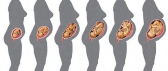
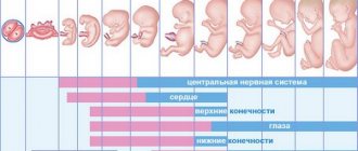
The location of the baby in a woman’s uterus begins to matter only after 32 weeks of gestation, when the baby is already quite large and it becomes more and more difficult for him to turn over in the stomach . Until this period, the child can spin in the womb as he pleases.
Head presentation of the fetus is the most common and natural position of the fetus in the stomach of a pregnant woman. The baby is in the womb with its head down at the entrance to the pelvis, the legs, accordingly, are located at the top of the abdomen. In 96–98% of cases, the child is in a cephalic position before labor, which is the most favorable for its good course. Other types of presentation are pathological and often childbirth with them may require surgical intervention. In addition to the favorable one, there are also such abnormal types of position of the child in the uterus before birth as: pelvic, transverse and oblique.
All types of fetal presentation except cephalic presentation are considered pathological
Variants of cephalic presentation of the fetus:
- occipital;
- anterior cephalic (parietal);
- frontal;
- facial.
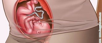
The preferred position for the birth of a child is cephalic occipital presentation . The baby lies in the tummy with the head tilted towards the chest, down. The baby's face is mainly turned towards the mother's spine. Thus, the baby will go out through the narrowest place of the head - the back of the head. The occipital position of the fetus at birth is the least traumatic for the child and for the woman; childbirth usually takes place without severe damage or complications. The fetus moves along the birth canal with the back of the head forward, with the neck bent and the chin pressed to the chest.
Occipital presentation is the most favorable position of the fetus before birth
The anterocephalic or parietal position of the fetus is noted when the baby is facing the genital tract with a large fontanelle, the head is not tilted towards the chest, but is in a straight position. Going through the cervix and vagina with such a presentation will initially be crown. Delivery is possible both with the help of surgical intervention and by the woman’s own efforts. But in both situations, mandatory monitoring of the baby’s condition is necessary to avoid a lack of oxygen. Childbirth with an anterior cephalic position of the baby is often protracted and traumatic for the child or mother.
The anterior cephalic position of the fetus during childbirth can be traumatic for mother and baby
Frontal presentation of the fetus is a rare, but at the same time the most dangerous position of the child before childbirth . Occurs in only 1–2% of pregnancies. The baby's neck straightens and the baby's forehead becomes directed towards the birth canal. The fetus, in the event of birth, will have to pass through the genitals at the widest point of the head. The birth of a baby in a natural way with this presentation is unacceptable.
With frontal presentation, delivery is always performed through surgery.
Facial presentation is observed even less frequently than frontal presentation - in only 0.3% of all pregnancies. With this position, the baby's head is strongly tilted back and the back of the head is almost pressed to the back. In the case of childbirth, the baby moves along the birth canal face forward, and the nose and chin become the leading point. Childbirth can be either natural or through surgery. In each case, an individual decision is made regarding the type of birth, depending on many factors: the parameters of the woman’s pelvis, the weight of the child, the intensity of contractions. Despite all the circumstances of pregnancy, the birth of a baby naturally in the frontal position is almost as dangerous and traumatic as in the frontal position. Injuries to the child's spine are often observed. Often presentation changes to facial from frontal at the time of birth. Less commonly, facial position is diagnosed by ultrasound some time before birth. This type of pathology most often affects multiparous women.
Frontal presentation of the fetus is a 100% indication for cesarean section
What are the reasons for fetal malposition?
Sometimes gynecologists diagnose pregnant women not with a cephalic presentation of the fetus, but with a breech presentation. That is, the baby is not placed in the uterus upside down, but sits in the mother’s pelvic bowl with his legs or butt. The following factors provoke the child’s incorrect position:
- mother's narrow hips;
- myoma;
- polyhydramnios, which increases fetal activity;
- oligohydramnios, which interferes with the baby’s mobility;
- placenta previa;
- abnormal structure of the uterine walls;
- low contractility of the uterine muscles;
- genetic predisposition;
- constant compression of the abdomen during daytime rest and night sleep;
- wearing uncomfortable and tight clothing.
If the child is in a pelvic or transverse position, the gynecologist decides which method of delivery will be used. With pelvic placement of the fetus, both normal childbirth and surgical intervention are allowed. Obstetricians perform a caesarean section if the woman in labor has too narrow hips or if the baby is large. In case of transverse presentation, when the baby is located horizontally in the uterus, obstetrics are carried out exclusively by cesarean section. Natural childbirth is contraindicated.
Causes of pathological types of cephalic presentation on the part of the mother and fetus
Causes of pathological presentation of the fetus on the part of the woman:
- narrow pelvis of a pregnant woman;
- too weak tone of the uterus, irregular compression of its sides;
- excess amniotic fluid;
- second or more births;
- multiple pregnancy;
- uterine fibroids;
- saddle or bicornuate uterus;
- the presence of a septum in the body of the uterus.
Causes of pathological position on the part of the fetus:
- tumor of the baby’s neck or thyroid gland;
- too small, or vice versa, very large head, which is associated with diseases.
Why is the baby positioned incorrectly?
The causes of breech presentation are not well understood, but factors that contribute to this type of presentation are identified.
Factors:
maternal factors:
- abnormalities of the uterus;
- uterine tumors;
- operated uterus;
- complicated obstetric history;
- tumors of the pelvic bones;
- narrow pelvis;
- decreased or increased uterine tone;
- a woman after a large number of pregnancies.
fruit factors:
- intrauterine growth retardation;
- congenital fetal anomalies (hydrocephalus);
- abnormal position of the fetus;
- premature baby;
- multiple births;
- immaturity of the vestibular apparatus of the fetus;
- decreased motor activity;
- decreased muscle tone;
placental factors:
- placenta previa;
- localization of the placenta in the fundus;
- short umbilical cord;
- umbilical cord nodes;
- polyhydramnios;
- oligohydramnios.
Diagnosis of cephalic presentation
A specialist can manually assess the position of the child after 22 weeks of an interesting position. However, before the 32nd week of pregnancy, the baby may change its position several more times. After 32–35 weeks, the baby is most often breech, which will persist until delivery.
In 95% of pregnancies after 35 weeks, the position of the baby in the uterus remains unchanged.
In order to understand the presentation of the baby, the obstetrician-gynecologist, with an open palm, tries to feel the baby’s head in the lower abdomen (the junction of the lateral pelvic bones). When the baby is positioned head down, it is palpated and determined to be a dense, rounded part. In addition, the doctor uses a technique called voting. If the palpable part of the fetus is lightly moved a couple of times, the head wanders (oscillates) in the amniotic fluid. The baby's heart, when positioned head down, usually beats just below the mother's navel, which the doctor can also find out using a fetal doppler. The gynecologist can clarify the type of position of the baby using a vaginal examination.
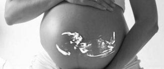
If there are any doubts about the position of the baby in the womb, it is advisable for the pregnant woman to undergo another ultrasound.
Mommy can assess the location of her baby on her own. To do this, she should lie on her back with her legs bent at the knees. Using the fingers of one hand, try to gently feel the head in the lower abdomen. If this succeeds, the position is in the head position. It is impossible to find out the type of head position at home.
Using an external examination, the doctor can determine fetal presentation
Exercises to correct abnormal types of baby presentation
If, after thirty-two weeks, a pregnant woman is found to have an abnormal position of the baby, doctors may recommend performing useful exercises that will encourage the fetus to assume a normal position for childbirth. However, it is worth remembering that sometimes there are individual characteristics of the female body or child that can prevent the baby from turning into the correct position, despite the quality of the exercises performed by the woman.
There are also restrictions on performing gymnastics by a pregnant woman:
- risk of untimely birth;
- placenta previa.
If there are no restrictions, a woman can try doing simple exercises:
- Lie on your side on a hard surface. Lie down for 12–15 minutes on one side, then turn over to the opposite side and lie down for the same amount of time. You should roll over from one side to the other at least three times in one approach. It is worth performing these manipulations two to three times a day. The effectiveness of simple actions is often high and the result appears already in the first 5–7 days.
- Lie on your back, place blankets under your legs and lower back so that your legs are 25–30 cm higher than your head. You need to lie in this position for 20–25 minutes, 1–2 times a day.
- Take a knee-elbow position on the floor and stay in this position for 20–25 minutes. The exercise should be performed 3-4 times a day.
It is recommended to stay in this position for at least 20 minutes per approach. - Exercise “Cat”, for which you initially also need to take a knee-elbow position. While in the starting position, bend your back up in the lumbar region, then straighten your back without bending down. The exercise must be performed 15–20 times in one approach, 2–3 times a day. “Kitty” is useful for all pregnant women who have no contraindications; when performing this exercise, the uterus is saturated with oxygen.
- Stay in a standing position or slightly leaning forward for longer than usual.
- Try to sleep on the side where your baby's head is.
- Swim on your stomach and back.
Swimming helps turn the fetus head down - Starting position: standing, feet shoulder-width apart, hands on the belt. Perform slow bends forward and backward. Inhale while standing, exhale while bending over. It is recommended to perform 10–20 smooth bends in one approach.
- The starting position is the same. Perform smooth bends to the side. As you inhale, be in a horizontal position and raise your arms up; as you exhale, lower your arms and tilt to one side. It is worth performing 10 bends in each direction at a time.
When performing any of the exercises, a pregnant woman should not experience discomfort, severe physical exertion or pain. To achieve the best effect, it is recommended to perform gymnastics no earlier than 2-3 hours after eating. Movements help increase the tone of the uterus and stimulate the baby's motor activity.
People say that you can help your baby roll over using a headphone with music or a light source placed on the lower abdomen. They say that the baby, even in the womb, begins to pick up sounds and changes in lighting and, out of curiosity, can turn towards an external stimulus.
If the mother’s efforts were not in vain and the child, with the help of physical exercises, nevertheless took the necessary position, it is worth helping him remain in a natural presentation. First of all, for this you need to wear a bandage. The bandage should be put on while lying down and worn for at least half a day. It also makes sense to do a couple of simple exercises:
- sit on the floor, bend your knees and bring your feet together. Press your knees to the floor as much as possible and sit in this position for 5-15 minutes;
- sit on a chair, press your feet to the floor. Slowly raise your bent legs up 20–30 cm, and then lower them to the starting position. It is recommended to perform 20–30 repetitions in one approach.
It also happens that the baby independently turns over into a natural position for childbirth in the last weeks of pregnancy. Still, nature takes its toll and there is no need to panic ahead of time.
That's what happened to my friend. After 32 weeks, she was informed that the baby was in a breech position. She began to do physical exercises to make the child turn over, but they did not bring results. And a week later, after the girl stopped doing gymnastics, at the next ultrasound she was pleased that the fetus had turned head down.
External obstetric inversion
Another way to turn the baby into the desired presentation is an external obstetric inversion. It is usually performed after the 35th week of pregnancy by experienced specialists strictly in a hospital setting. The manipulation is also carried out under the control of ultrasound and CTG.
Before starting the inversion procedure, the doctor monitors the position of the fetus using ultrasound and performs CTG, so the doctor has a good idea of what is happening inside. After this, using gentle hand movements on the pregnant woman’s belly, the doctor tries to turn the baby head down. Typically the manipulation lasts from several seconds to several minutes. After the revolution, the condition of the fetus is again assessed using an ultrasound machine and CTG. Then the woman goes home.
Some women are afraid of performing an external rotation, assuming that the doctor’s actions may injure the child. But that's not true. The fetus in the womb is in a state of hydro-weightlessness and cannot be injured by the experienced actions of a doctor.
Obstetric fetal inversion does have small risks, which are discussed with the patient before the manipulation, but the risk from external inversion is very small and is not comparable to the danger of giving birth to a child in an incorrect presentation or cesarean section. All over the world, there is no knowledge of the real complications that the procedure of obstetric fetal inversion would bring.
In approximately 35–40%, external rotation cannot be carried out . This is mainly due to the presence of contraindications to this manipulation. Less often, there are cases when the fruit itself could not be turned over. It depends on the stage of pregnancy. The higher the gestational age, the more difficult it is to successfully perform an external obstetric rotation of the baby.
Video: external obstetric inversion
Features of childbirth with facial, frontal and anterior cephalic types of presentation
The birth of a child in the facial, frontal and anterior cephalic (parietal) position is pathological. In the face presentation, the fetal head enters the small pelvis with the largest size of 32–33 cm, the leading part is the chin. In a presentation with the forehead forward, the largest size of the head passing through the birth canal is 39–41 cm, and the leading part is the eyebrow. With anterior cephalic presentation, the maximum circumference of the head passing through the genitals is 34–35 cm, and the leading point is the crown.
In many cases, during labor, the type of abnormal presentation may change. Thus, the parietal position, when the head is tilted forward, passes into the occipital position, and when extended, into the frontal position.
The anterior cephalic position of the baby is often accompanied by untimely rupture of amniotic fluid. The decision regarding the type and strategy of childbirth is made individually in each case; independent childbirth and caesarean section are possible. A woman will be able to give birth on her own if the fetus is small and the head can pass through the genital tract with the crown. During normal childbirth, the initial periods of birth last longer than with occipital presentation, and the likelihood of trauma to the woman’s genital organs, as well as the likelihood of trauma to the child and its hypoxia, increases. There are cases when the baby’s head enters the pelvis and after this the woman in labor experiences weakness in labor. In such a situation, specialists remove the fetus using obstetric forceps or a vacuum extractor (a device that pulls the baby out of the genitals using rarefied air).
During prolonged labor and weak labor, obstetricians-gynecologists use a vacuum extractor to extract the baby.
The small pelvis of the woman in labor, the large head of the baby, weak contractions, post-term pregnancy - serve as indications for the use of an operative method of delivery.
Particularly dangerous for childbirth is frontal presentation. The baby's head lies at the entrance to the genital tract at its largest size. Therefore, independent childbirth in this position of the child is practically impossible, and if it takes place, it is with severe damage to the child and mother. A woman may experience a rupture of the perineum or uterus. Untimely discharge of amniotic fluid is observed, which can lead to oxygen starvation or infection of the fetus. The duration of labor increases. The only possibility of independent birth is to correct the position of the fetus to the parietal or facial position, but it will still not be possible to avoid damage to the child.
Due to the danger of serious consequences of natural childbirth, in almost 100% of cases, childbirth with the frontal position of the fetus is carried out using surgery. The main thing is to have time to carry out surgical intervention before the head enters the pelvis. If the head has entered the small pelvis and remains in a static state for a long time, signs of fetal hypoxia are noticed - specialists usually resort to the help of obstetric forceps. If the forceps do not help and the fetus cannot be extracted, it is necessary to perform an operation that destroys the fetus in order to save the life of at least the woman in labor.
With a facial presentation of the fetus, prenatal rupture of amniotic fluid and oxygen starvation of the fetus may also occur, and umbilical cord prolapse may occur. The duration of birth increases by almost one and a half times when compared with childbirth in a favorable position. A significant factor is keeping the fetus in the posterior position, otherwise a transition to the frontal position is possible and independent childbirth will become impossible. If the type of presentation has changed to frontal, an immediate cesarean section is required, or, if it is too late and the head is stuck, specialists perform a fetal-destroying operation.
If the birth goes according to plan, the child’s skull needs to adapt to the birth canal and change its shape, which is why a birth tumor is formed. The parietal bones may also overlap each other. In both situations, the baby’s head takes on a normal shape for some time after birth.
In 92–95% of births with the fetus in the facial position, they occur naturally; in other situations, a cesarean section is necessary.
How often is a caesarean section performed?
With the longitudinal position of the fetus and cephalic presentation, cesarean section is rarely done. According to various sources, about 95% of children are born in the occipital presentation, about 1% in the anterior cephalic presentation, up to 1% in the frontal presentation, and less than 0.5% in the facial presentation. Indications for cesarean section are extreme degrees of extension presentation (facial, less often frontal, even less often anterocephalic).
Operation caesarean section
Measures to prevent abnormal fetal positions
In fact, there are no special specific measures to prevent the risks of malpresentation of the fetus. Everything is natural, and the correct position of the child in the womb is inherent in nature; you just need to ensure the normal course of pregnancy throughout its entire period.
It is advisable for a pregnant woman to maintain a favorable psychological mood, eat a nutritious and varied diet, and observe a work and rest schedule. You shouldn't overeat either, especially in the last weeks of pregnancy. After all, a large and post-term fetus can be a risk factor for malpresentation. You should also strictly follow your doctor’s recommendations and take vitamins or medications as prescribed by specialists. If necessary, it is possible to perform special gymnastic exercises suggested by the obstetrician-gynecologist.
Communicate more with your baby, ask him to take the correct position in his tummy - this will help both you and the baby tune in to a positive wave before giving birth.
How does pregnancy proceed during breech presentation?
The diagnosis of breech presentation is made after examinations:
- external obstetric examination according to Leopold:
- determine the position of the fundus of the uterus (above normal);
- when palpated above the entrance to the pelvis, a large, soft, irregularly shaped presenting part of the fetus is identified, which is not capable of changing position; in the fundus of the uterus there is a dense and rounded head of the fetus, which can turn left and right;
- the fetal heartbeat can be felt above the navel;
- Ultrasound of the fetus at 32-34 weeks of pregnancy:
An ultrasound in the third trimester determines the position of the fetal head. To do this, measure the angle between the back of the head and the baby’s spine. The greater the angle, the less likely it is to give birth spontaneously.
There are 4 options for the position of the fetal head during breech presentation:
- the head is bent;
- the head is slightly extended (military pose);
- the head is moderately extended;
- excessive extension of the head (“looks at the stars”).
- Fetal ECG;
- amnioscopy;
- vaginal examination during childbirth with sufficient dilatation of the cervix and absence of amniotic sac.
With breech presentation during pregnancy, the following complications may occur:
- gestosis;
- umbilical cord entanglement;
- threat of miscarriage;
- intrauterine growth retardation.
If a pregnant woman is diagnosed with a breech presentation of the fetus, then wait until the 28th week of pregnancy. Then, if the situation has not changed, then they prevent and correct the breech presentation to the cephalic one.
Starting from week 32, 37 women are recommended to perform special gymnastic exercises.
If attempts to change the position of the fetus fail, the pregnant woman at 37-38 weeks is transferred to a hospital and tactics for managing future births are chosen.