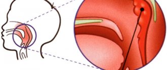
The normal course of labor is characterized by progressive effacement, dilatation of the cervix, descent of the presenting part of the fetus, and advancement of the fetus along the birth canal. To correctly assess the clinical course of labor, it is necessary to understand its biomechanism, i.e. the order of the main movements of the fetus as it passes through the birth canal.
- Biomechanism of childbirth
- Birth tumor
- Periods of childbirth. Management of childbirth
- Vaginal delivery
- Episiotomy
Fetal presentation - what the expectant mother needs to know
Many mothers expecting a baby for the first time do not always understand what they are talking about when the doctor talks about the presentation and position of the fetus. Let's figure out the baby's orientation in space!
Until approximately the 30th week, the baby moves freely in the uterus. However, by 35 weeks it is too large and too crowded in the uterus for it to turn over easily. Therefore, the obstetrician-gynecologist tries to determine the position in which the child finds himself - the birth strategy depends on this.
Baby's position
The position of the child is determined relative to the position of the mother's body.
- Longitudinal position - the child is located along the mother’s body, the “head-buttocks” axis of the fetus coincides with the mother’s. This is the best orientation for natural childbirth.
- Transverse position – when the baby is positioned across the mother’s belly and is “stuck” in this position. Natural delivery in this case is impossible and this position of the fetus becomes an indication for cesarean section. However, before making such a decision, the doctor can wait until the amniotic fluid is released: the uterus becomes more spacious and the child can turn his head “towards the exit.”
- Finally, the oblique position of the fetus is all intermediate options between longitudinal and transverse. In this case, natural childbirth is also difficult, and the obstetrician will recommend doing special exercises to make the fetal position more successful.
Head Configuration
Head configuration represents changes in its shape due to external pressure. A certain configuration of the head may occur before birth, as a result of Braxton-Hicks contractions. The most common form of head configuration is that the parietal bones overlap each other. The head configuration results in a decrease in the minor oblique and an increase in the major oblique diameter of the head. These changes are very important for narrowing of the pelvis and asynclitic insertion of the head. In these circumstances, the ability of the head to change is of great importance in spontaneous vaginal delivery and operative vaginal delivery.
Presentation of the baby
In addition to the position of the child, it is important to determine his presentation - he is located with his head or buttocks down. Breech presentation is quite rare (approximately 3% of women in labor) and is not an indication for cesarean section. However, managing such a birth requires the doctor to have great knowledge, skills and experience, as well as preparedness for emergency situations (when a caesarean section is still required).
Head presentation
Even if the baby is positioned head down, different parts of the head may be directed towards the cervix, and accordingly, the neck can be arched differently (more or less).
- The most favorable situation is if the child approaches the birth canal with the back of the head, facing forward - this is how 90% of all newborns are positioned. In this case, childbirth is easy and without complications.
- It happens that the baby's face is turned towards the mother's back, the so-called posterior view of the occipital preposition. Childbirth in this case lasts a little longer and requires more attention from the obstetrician.
- The situation is somewhat more difficult with anterior cephalic presentation, when the baby’s head is directed into the birth canal exactly in the center, approximately by the fontanel located at the junction of the frontal and parietal bones.
- A greater bend in the neck will force the baby to rest his forehead on the cervix, increasing the likelihood of possible complications during childbirth.
- Finally, the most dangerous of the cephalic presentations is the facial presentation. The child’s neck is fully extended, we can say that the baby is “not grouped” - in this case, natural childbirth is fraught with the danger of serious injury to the child’s neck, and, most likely, the doctor will raise the question of a caesarean section.
Breech presentation
If the baby is positioned head up before birth, then options are possible in this case as well.
- The child is positioned towards the birth canal with the buttocks, the legs are extended and pressed to the body. This is the most favorable option for natural childbirth.
- The legs extended towards the cervix significantly complicate the process of labor - the leg may fall out when it gets stuck in the birth canal during pushing.
- Finally, the most difficult option is mixed presentation, when the baby’s knees or buttocks with crossed legs are directed towards the birth canal.
Prolapse of the fetal head: how it happens
The most important harbinger of childbirth is prolapse of the fetal head, which experts call fetal formation or uterine prolapse. For each pregnant woman it occurs at a different time, but the process itself and the different sensations are the same for almost everyone. A drooping abdomen immediately before childbirth should not greatly frighten a pregnant woman.
Prolapse of the fetal head: what happens in the body?
In the last months of pregnancy, the belly begins to drop because the baby is preparing to be born, so very serious changes are currently taking place in the body of a pregnant woman, which directly affect her well-being. During the descent of the fetal head immediately before childbirth, the uterus begins to change its position, as well as the fetus that is in it. During this period, the following changes occur in the pregnant woman’s body:
- The fetus begins to move down the uterus with the presenting part (this is the part of the body that is the first to be born: usually the head appears first);
— The uterus begins to fall down from its usual position by about 5 centimeters;
— When the fetal head descends, the baby takes the most comfortable place in the pelvis, where it will remain until labor begins. This arrangement is called a tuck and is a bit like preparing an athlete to start a race;
- At this moment, the uterus no longer puts pressure on the diaphragm, which finally has the opportunity to fully expand;
— Organs located in the gastrointestinal tract no longer feel pressure from the uterus and fall into place;
— The heavy fetus puts pressure on the pelvic bones and legs.
All the changes that occur during this period affect the general condition of the woman, who must know all the signs and be prepared for them in advance.
The main signs of fetal descent
In every pregnant woman, the prolapse of the fetal head before the onset of labor will be characterized by general signs, which can be used to determine readiness for future contractions. Many expectant mothers almost accurately guess this phenomenon, since it is impossible to feel these signs. The main signs of fetal descent can be positive, external and not very pleasant.
Positive signs of head prolapse
- It becomes much easier for a woman to breathe, shortness of breath goes away: the diaphragm no longer feels the pressure of the uterus, as a result of this the pregnant woman can breathe freely;
— Severe pain in the ribs disappears;
— The child’s movements are now practically painless;
— When the fetal head drops, heartburn, belching, nausea and heaviness in the stomach, which were the woman’s companions throughout almost the entire pregnancy, disappear, since there is no more pressure on the organs of the gastrointestinal tract, and they are able to work normally;
Unpleasant signs
- Constipation may occur;
— There is a frequent urge to go to the toilet, because during this period the uterus puts pressure on the rectum and bladder. This is also explained by the fact that before childbirth the body begins to cleanse itself of all unnecessary substances;
— When sitting and walking, a pregnant woman feels discomfort, this is due to the fact that the fetus is large in size, and it begins to put pressure on the pelvic bones;
— Painful sensations may appear in the pelvis, lower extremities and lower back: this can be explained by the fact that the child begins to put pressure on the nerve endings with his head.
Turn the child to the desired side
Surprisingly, gymnastics can affect a child’s position. An obstetrician-gynecologist can recommend exercises that are suitable specifically for your case, but in general, a complex of special gymnastics looks like this:
- Place your feet shoulder-width apart, arms hanging freely. As you inhale, raise your straight arms to shoulder level, stand on your toes and arch your back, taking a deep breath, and exhale to return to the starting position. Repeat 4-5 times.
- Lie on your side, bend your legs towards your stomach and lie in a comfortable position for 5-10 minutes. Then roll over your back onto the other side, slightly stretching the oblique abdominal muscles, as if “stretching” sideways, and lie down some more. Repeat 5-6 times.
- Lying on your back, bend your knees, rest your feet on the floor. Inhale, lift your pelvis off the floor, and exhale, lower it. Repeat 5-7 times.
- If the last part of the exercise is difficult to do, you can place a couple of pillows under your buttocks so that your pelvis is 30 cm above shoulder level, and lie in this position for about 10 minutes.
Exercises should be done before meals, the complex can be repeated several times a day.
Remember that any exercises can only be done after consulting with your doctor, since even such a harmless, at first glance, complex has a number of contraindications: gestosis, the threat of premature birth, a scar on the uterus left after a cesarean section, placenta previa.
Maybe the doctor will do it himself?
Previously, obstetricians actively tried to turn the baby with their hands, pressing on the mother’s stomach. Today, such methods are not practiced - the risk of complications is too great: placental abruption and premature birth.
No less dangerous is obstetric intervention carried out directly during the birth process, the so-called “turn on the leg.” In this case, when the cervix is fully dilated, the doctor inserts his hand into the uterus of the woman giving birth and grabs the baby’s leg, while with the other hand, through the abdominal wall, trying to fix the baby’s head at the top. Then the baby is actually pulled outward by the legs - the birth proceeds in the same way as with a foot presentation. Today, this method is used only in exceptional cases, during the birth of twins, when after the birth of the first baby the second suddenly took a transverse position.
Vaginal delivery
When the fetus begins to be born (“head tapping”), the maternity unit staff observes asepsis and puts on sterile medical clothing, masks and gloves (prevention of maternal and fetal infection and self-protection). The necessary tools include two clamps, scissors and a suction device.
During the birth of the fetal head, techniques are used aimed at protecting the perineum from trauma (regulation of pushing, maintaining intervals between pushing, careful, controlled removal of the head). With one hand, the perineum is supported (“removed” from the head) and gentle pressure is applied to the fetal chin in an upward direction, and with the second, gentle pressure is applied (flexion of the head) to prevent its premature extension and injury to the perineum, as well as massage of the labia before birth (“teruption”) ") heads.
Immediately after the birth of the head (before the birth of the shoulders), the contents of the upper respiratory tract of the fetus are suctioned. If there is meconium in the amniotic fluid, the contents of the newborn's oropharynx and nasopharynx are carefully suctioned using a special catheter before the birth of the shoulders, when the newborn's chest is still compressed by the mother's birth canal and he cannot take his first breath (preventing meconium aspiration).
After complete suction of the mucus, check for the presence of entanglement of the umbilical cord around the fetal neck. If such a condition exists, and the doctor is confident that labor will end soon, the umbilical cord is cut between two clamps. If labor may be complicated by shoulder dystocia (difficult delivery of the shoulder girdle, for example, in the case of fetal macrosomia), efforts are made to hasten the birth of a fetus with an intact umbilical cord.
After external rotation of the head towards the mother's thigh, the birth of the anterior shoulder is assisted by applying downward pressure on the head with the palms. Once the anterior shoulder is visualized, pressure is applied to the head in the opposite direction (upward) to assist in the delivery of the fetal posterior shoulder. After the head and shoulders are born, light traction is applied to speed up the birth of the rest of the fetal body. After this, the umbilical cord is crossed between two clamps and the newborn is handed over to the mother or midwife, and, if necessary, to a pediatric neonatologist who is in the delivery room.
What it is
Every pregnant patient knows that this is a cephalic presentation of the fetus. This indicates the location of the fetus in the uterus with its head towards the entrance.
Taking into account which part of the head is turned towards the neck, the main types of presentation are distinguished. Everyone influences how the birth will take place, since situations can be unpredictable.
There are times when the use of obstetric forceps or cesarean section will be necessary. It is important that the specialist reacts in a timely manner and makes the right decision.
Classification
Head presentation of the fetus during pregnancy has a certain classification. It is determined by a specialist in the last stages of gestation. Gynecologists highlight the main characteristics of the location, which are very important:
- view - how the child is turned;
- position – direction to the right or left (for example, the first indicates a turn to the right).
Changes may occur at any time. The birth of a small life is unpredictable.
Occipital
This type is considered the most successful. The head is tilted down towards the chest, and the face is turned towards the spine of the expectant mother.
The baby comes into the world through the cervix with the back of the head. This is the narrowest part. This position is safe and has little risk of injury. The process proceeds largely without damage.
Anterocephalic
Anterior cephalic presentation is also called parietal presentation. In this case, the fetus is turned towards the genital tract by a large fontanel.
His head is not tilted to his chest enough and is held straight. Initially, the crown of the head will emerge through the birth canal.
The woman in labor can cope on her own or will need surgical intervention. It is necessary to strictly control this process, since at any moment there may not be enough oxygen and a threat appears.
Facial
It is observed very rarely. This position is accompanied by a strong deviation of the child’s skull back, and the back of the head is pressed to the back. Exit to the light is done face forward, nose and chin.
The mother has a chance to give birth on her own, but if necessary, a caesarean section will be performed. The gynecologist makes a decision individually, taking into account the labor activity of each patient.
This is influenced by the following factors:
Considering all the data, facial presentation is dangerous. It often causes injuries to the newborn's spine. Mainly observed during repeated and multiple gestations.
Frontal
Frontal cephalic presentation of the fetus is extremely rare and very dangerous. Occurs in 1-2% of expectant mothers. At the same time, the child’s neck straightens, and he is directed towards the exit by the frontal part.
The danger is that the passage through the birth canal is carried out by the widest parts of the head. In this case, natural delivery is prohibited, so only cesarean section is performed.
The stomach has dropped - a sign that will show when and how long to give birth
Pregnant women always pay special attention to changes in their bodies. Some begin to worry that the belly has dropped, others are worried that at 38-39 weeks of pregnancy this has not yet happened.
As a rule, abdominal prolapse is one of the harbingers of childbirth. In connection with this, the question arises, how many days or weeks before childbirth does the belly drop? The period is different for each woman.
There are many reasons why this does not happen. But this does not mean that a woman’s body is not preparing for childbirth. For obstetricians, the concept of “abdominal prolapse” is not only a harbinger of childbirth, but also an indicator of the proportionality of the pelvic ring to the parameters of the baby’s head.
When the belly drops during pregnancy: a little physiology
After the 37th week of pregnancy, a “dominant of labor” begins to form in the cerebral cortex of a woman. From this moment the body begins to prepare for the birth process.
https://www.youtube.com/watch?v=l2jsZ4jc0WQ
The hormone relaxin is produced, which helps relax connective tissue and tendons. Under the influence of this hormone, the articular joints of the woman’s pelvic ring begin to “diverge” a little, this process is especially pronounced in the pubic symphysis. Thanks to these processes, the woman's pelvis adapts to the upcoming birth.
Many women, already at 35-36 weeks, are interested in how long before giving birth the belly drops.
By the beginning of the 37th week, the lower segment of the uterus is formed.
This area anatomically corresponds to the isthmus of the uterus, but it is in the last weeks of pregnancy that it begins to significantly increase in size. Due to this, the lower part of the uterus increases, as a result of which the fetal head descends freely and is fixed to the pelvic bones. Such a change in the position of the fetus also leads to changes in the position of the uterus: its fundus drops significantly.
After the head lowers to the entrance to the pelvis, the position of the woman’s center of gravity changes. The woman develops a gait that obstetricians call a “proud walk.” Since the main load falls on the lower back, the woman walks with a straight back, raising her head slightly, often holding her lower back with her hand.
conclusions
A drooping abdomen before childbirth is an important sign that a woman’s body is ready for childbirth. But obstetricians attach importance to this sign for another reason: the stomach drops if the sizes of the fetal head and the pelvic ring are comparable.
Thus, this sign should be treated with increased attention, since it reflects many physiological processes before childbirth: the formation of the lower segment of the uterus, fixation of the head to the pelvic bones, adaptation of the pelvic ring, correct position of the head.


