The spleen is an organ of our body that is not fully studied. However, the established functions assigned to this body are quite extensive. Among the functional responsibilities of the spleen are the functions of hematopoiesis, the fight against various viral and bacterial pathogens.
The spleen acts as a kind of filtration organ of the blood from pathogens. The spleen contains a large supply of red blood cells - erythrocytes, which can be used to restore blood volume during sudden blood loss.
Preparation for ultrasound of the spleen
It is necessary to prepare for an ultrasound of the spleen in the same way as for a similar examination of the abdominal cavity.
Before the examination, do not take food or water for 8 hours. Children, if conditions permit, should refrain from eating and drinking water for 3 hours before the test. It is advisable to stop taking foods that promote gas formation (sweets, legumes, cabbage, wheat bread, baked goods, milk, etc.) 2-3 days before the ultrasound. Preparation for an ultrasound involves taking enterosorbents, such as lactofiltrum, activated carbon, smecta for several days before the ultrasound. These drugs are taken one tablet two to three times a day. Taking enterosorbents is necessary in order to completely avoid gas formation, which can greatly complicate this study.
Ultrasound of the spleen
The ultrasound of the spleen itself lasts five to ten minutes. During the ultrasound, the patient does not feel any discomfort, and there are no contraindications to ultrasound examination. First, the ultrasound doctor scans the organ in a position where the patient is lying on his back, and then lying on his right side.
Kidneys of different sizes in a child
If an ultrasound reveals that a child has one kidney lower than the other, there is no need to worry immediately, since in adults, the right kidney is normally located half a vertebra lower than the left. Their upper pole is recorded on ultrasound at the level of the twelfth thoracic or first lumbar vertebra.
It is much more dangerous if they are located higher in the chest area (diaphragmatic hernia) or lower at the level of 1–5 lumbar vertebrae (lumbar, iliac pelvic dystopia). This arrangement causes severe pain in the newborn when he strains to cry. In addition, dystopic organs are more often susceptible to the development of pyelonephritis, hydronephrosis, and the formation of stones.
As for the asymmetry of the kidneys in a child, in adults, healthy people, the sizes of the kidneys may also differ. The left one is usually slightly larger than the right one. Significant changes in size, their inconsistency with the baby’s age, weight, and structural changes in them require attention.
Symptoms
It is impossible to quickly guess that a child has an enlarged spleen. Usually the process of splenomegaly itself does not cause any clinical symptoms. The child may be tormented by manifestations of other diseases that were primary in relation to the enlarged spleen. Typically, parents learn about splenomegaly only during an examination:
- The inflammatory process in the spleen is characterized by such manifestations as frequent and fairly prolonged diarrhea, slight nausea and occasional vomiting, pain under the ribs, and increased body temperature.
- Non-inflammatory processes in the spleen rarely cause pain on palpation. The temperature also usually remains normal. The skin in pathologies associated with an enlarged spleen may be pale, the child may become more tired and apathetic. Increased sweating may occur at night.
Pathological foci
The splenic parenchyma looks heterogeneous on ultrasound. Foci of different sizes, contours and density indicate a specific disease. A dark lesion with smooth contours and uniform echogenicity indicates a benign splenic cyst.
Heterogeneous lesions with unclear contours should alert the researcher. This could be a life-threatening tumor (lymphoma) or an acute purulent disease - an abscess. Light, blurry spots will suggest foci of metastasis.
An increased size of an organ with a homogeneous structure and rounded edges will indicate an inflammatory process. If dark, small lesions appear against this background, then the disease has become chronic, and foci of dead cells (necrosis) have appeared in the parenchyma.
In the future, these “scars” in the tissues will become denser and remain light, uneven spots for life. Ultrasound gives a different picture of tissue necrosis due to vascular thrombosis. A wedge-shaped area of low echogenicity (dark spot) will appear on the screen. Its structure will be homogeneous, and its contours will be blurred.
With splenic abscesses, the echogenicity levels of the lesions will undergo changes depending on the stage of the process. Light spots gradually appear on the dark lesion, and then a light capsule with a dark spot in the middle is formed.
Ultrasound can detect parenchymal rupture. The following picture is determined:
- contour discontinuity;
- the presence of layers - internal and external;
- dark blood stains between layers.
Hemorrhage is defined as dark areas. As they dissolve, the spots lighten and then disappear completely.
How to prepare?
Correct interpretation is possible with a high-quality ultrasound of the spleen. This requires proper preparation. Three days before the examination, you should not eat foods that contribute to gas formation: legumes, milk, rye bread, raw vegetables. It is also recommended to take sorbents and enzyme preparations that stimulate digestion (mezim, meteospasmil).
The procedure is performed in a certain body position. The patient takes a position on his side, the left cancer is raised behind the head. In a state of inspiration, a sensor through the intercostal space visualizes the state of the organ.
Important: you should not do an ultrasound of the spleen immediately after an endoscopic examination or diagnostic x-ray. This may skew the results
Preparing children for ultrasound diagnostics has its own particularities. Infants should not be fed before the procedure. Children from one to three years old should not eat for 4 hours before diagnosis, over three years old - 6 hours. You cannot drink for 1 hour.
Features of the structure of the spleen in adults
The spleen is a functional organ that is present in the body of every person from birth. She takes an active part in the processes of combating pathogenic pathogens that can disrupt the functioning of internal systems. The spleen is involved in the formation of immunity, the metabolism of lipids, proteins and carbohydrates, as well as in solving problems associated with the functioning of the hematopoietic system.
Any deviation in the functioning of an internal organ can result in serious problems for a person in terms of the function of the systems that are associated with it.
Anatomy and physiology
The spleen is located below the diaphragm, in the upper left part of the abdominal cavity, which is commonly called the left hypochondrium.
The internal organ is an unpaired lymphoid formation, which is located in the abdominal cavity. It can be found under the place where the diaphragm is located. The walls of the spleen are normally in contact with other organs. We are talking about the colon, stomach, kidney and pancreas.
The spleen is shaped like an elongated hemisphere, which is slightly flattened. The internal organ is covered by a connective tissue capsule characterized by a high level of density. Trabeculae extend from it directly into the parenchyma.
There is a diameter and a length at the spleen. They are felt by the doctor at the time of palpation of the internal organ. It itself is penetrated by vessels that are responsible for the deposition of blood.
Normal sizes
People who are concerned about their own health may be interested in the normal size of a healthy and diseased spleen. To find out whether a person has problems with this organ, it is necessary to conduct a comparative description of normal and current indicators that were clarified during diagnostic procedures.
The normal sizes of the spleen in children and adults are presented in the table.
Standards for adults During an ultrasound examination, the specialist takes into account several linear dimensions. The size of the spleen, presented as the norm in adults, is characterized by the following indicators: Length – 8-14 mm; Width – 5-7 mm; Thickness – 3-5 mm. As for the mass of the internal organ, in women it is in the range of 150-155 g
For men, the normal weight range is 192-200 g.
Norms for children and adolescents The norms for length, area, width and other indicators for children and adolescents are determined completely differently. A healthy spleen in a child corresponds to the following indicators: Length - 50-65 mm; Width – 17-25 mm. The above are the norms for a child who has reached one year of age. In a fifteen-year-old teenager, the organ has the following parameters: Length – 90-120 mm; Width – 34-49 mm. Medical reference books indicate more accurate standards for children and adolescents of different age groups.
Any deviation from the norm indicates that the spleen cannot cope with its basic functions due to damage by a pathological process. Even with mild ailment and discomfort in the area where the organ is located, it is strongly recommended to seek medical help to stop the progression of the disease.
Advertisements on NN.RU – For children
Factory Dastorg sells with free delivery in Nizhny Novgorod Dzerzhinsk. Banquette Litisia (Craft oak) Size (HxWxD). Price: 3,000 rub.
Children's lightweight sneakers, in good condition Price: 600 rub.
boots for teenagers, size 39, up to -25 Price: 1,500 rub.
The loft bed is in good condition. Mattress size 70*185. Under the bed there is a spacious built-in wardrobe for things, as well as a shelf. Price: 3,000 rub.
Coffee, cigarettes, food dyes quickly spoil the appearance of teeth; Plaque and tartar appear. So that the smile remains white and...
The ex-general director of Nizhny Novgorod Vodokanal, Alexander Popov, has been put on the wanted list due to fraud on an especially large scale.
Who said that the domestic auto industry is good for nothing? Just look at these strong Zhiguli cars cutting through.
Today in the village of Afonino there was a very serious accident: two cars flew into each other at speed. An eyewitness to the accident published photographs.
The spleen performs the most important function in the body - it regulates the hematopoietic system. An enlarged spleen indicates a pathological condition in the body and possible diseases. This condition is called splenomegaly.
Cyst on an organ: what to do, how to help with rupture
A splenic cyst in a child is usually detected completely by accident during an ultrasound scan. The treatment method for this pathology depends on the size of the formation. If a small cyst is detected, the child is monitored by specialists, and control examinations are done 2–3 times a year.
If a large or medium-sized cyst is detected, inflamed or ruptured, surgical intervention is performed. In this case, the cyst is removed, and in some cases the entire organ is removed.
An enlarged spleen in a child entails the destruction of blood cells. In turn, this condition can provoke rupture of the enlarged organ. How to determine pathology and what could be the cause? A fairly serious illness requires urgent medical attention.
Most often, a break does not occur suddenly. First, a hematoma forms, then under certain conditions it ruptures. In children, the organ is not sufficiently covered by the ribs, and therefore is poorly protected from external influences. In newborns, rupture occurs due to infectious diseases.
Characteristic symptoms of splenic rupture:
- in the upper part of the tummy, on the left, internal push;
- feeling of discomfort;
- a dull pain that gradually spreads throughout the abdomen.
Due to increased bleeding, other signs also appear:
- weakness and dizziness;
- child's pose: knees tucked to the stomach, on the side;
- a feeling of darkening and bright flashes in the eyes;
- nausea, vomiting, bloating, increased pain;
- when palpating the left side of the abdomen, pain radiates to the left shoulder blade.
What should parents do if they find signs of a rupture? This pathology requires surgical intervention, so you need to call an ambulance as soon as possible.
Prognosis after a child has had his spleen removed varies and depends entirely on the presence of concomitant diseases. Patients who do not have problems recover after 3 to 6 months. Children after splenectomy are prescribed a course of antibiotics to prevent possible infections, since the risk of their occurrence is high.
Diagnosis
The first thing that should be done is questioning the child’s parents in order to clarify previous diseases. Also take note and study diseases associated with the cardiovascular system, liver, and gall bladder. The patient is examined by palpation, the purpose of which is to determine the size of the spleen. Palpation is carried out when the patient is in a horizontal position with relaxed abdominal muscles.
During palpation , if you can feel the edge of the spleen, this indicates its increase in size. The patient should be sent for an ultrasound examination.
The purpose of ultrasound is to clarify the diagnosis. Also, to clarify the diagnosis and correctly determine the causes of an enlarged spleen, a variety of laboratory tests, blood, and urine are performed. With an enlarged organ, the chemical composition of the blood differs significantly from the norm. In any case, correct identification of the cause of the “disorder” of the spleen is half the way to providing the right help for the latter.
Normal on ultrasound
Immediately after the ultrasound of the spleen, its interpretation follows. A normal, healthy organ should be located in the upper part of the abdominal cavity, on the left side, and it is localized on the left, in the lower part of the diaphragm. The stomach should be located close to the middle of the organ, and the tail of the pancreas should be located near the middle of the gate. A grid of vessels may be visible in the goal area; this is not considered a deviation. The diameter of the splenic vein should not exceed half a centimeter. The parenchyma of a healthy spleen has a homogeneous fine-grained structure.
Not only the size of the organ is important, but also its echostructure, which must be homogeneous. There should be no inclusions in the image. Normally, the shape of the spleen resembles a crescent. If the organ has a heterogeneous structure (it is the heterogeneous structure that indicates benign tumors), the echogenicity is increased (in cancer of the blood it may not be increased, but splenomegaly is noted), the shape of the spleen is irregular - these are signs of the disease. Even small deviations from standard indicators are important and require consideration by a specialist.
For an adult
It is important that during ultrasound diagnostics three linear dimensions are taken into account. The scanning protocol must indicate specific numbers
There is not enough information about whether the spleen is enlarged. The normal dimensions of an organ for adults are as follows: the length of the organ is 8−14, the thickness is 3−5, the width is 5−7 cm. In women, the mass of a healthy organ is 150−152 g, in men - 192−200 g. The area of the maximum cut: normally it is 40-50 square meters. cm. It should be remembered that the size of the organ is individual for each person.
For children
The size of the spleen in children varies depending on age. For example, a one-year-old child’s spleen is relatively small: length – 50-65 mm, width – 17-25 mm. For a 15-year-old teenager, the acceptable indicators are: length - 90-120 mm, width - 34-49 mm. The table shows organ sizes for children of different ages:
This table will help determine whether the size of a child’s organ of a certain age category is acceptable. If they are not within normal limits, the following diseases are suspected:
- hematological syndrome;
- anemia;
- leukemia;
- Congenital heart defect;
- typhoid fever;
- tuberculosis.
Often an enlarged spleen in children indicates that the liver is unhealthy.
The spleen is a small internal organ and not everyone knows where it is. But in physical education classes, many of us often complained of pain in the left side that appeared during or immediately after running. This is the spleen, and it reacts this way to a sharp increase in blood volume.
Despite the fact that the absence of a spleen generally does not affect general well-being and a person without a spleen can live peacefully, its removal is not a disaster for the body. But the spleen is still a very important organ for many reasons - it fights diseases of the blood and bone marrow, participates in the formation of humoral immunity (when the body’s defense system produces special antibodies that fight infections) and cellular immunity (cellular immunity is responsible for resisting bacterial and viral infections). It is also involved in the metabolism of iron, lipids, proteins and carbohydrates.
The spleen is located in the left hypochondrium, between the diaphragm and the stomach at the level of the 9th to 11th rib. Unlike adults, in children the spleen is not completely covered by the ribs and is less protected from external influences (impacts).
The spleen develops throughout life and due to the proportional increase in the size of the spleen as the child grows, its shape does not actually change. But sometimes the spleen enlarges in both adults and children.
Enlarged "couple"
In most cases, enlargement of the liver, spleen and lymph nodes occurs simultaneously, since these organs are in direct relationship. An enlarged liver and spleen is the first sign of diseases of the hematopoietic organs and blood. In some cases, this symptom indicates the presence of infectious mononucleosis. Hepatolienal syndrome is a sign of chronic lymphocytic leukemia at the last stage.
Experts determine the cause of the simultaneous increase using a medical examination: blood biochemistry, X-ray studies, CT scans, scanning, ultrasound, MRI. These studies allow us to thoroughly examine the area of the spleen in children. There is no specific treatment for enlarged organs. Causes and treatment are closely related. After all, in order to successfully carry out treatment, it is necessary to correctly diagnose the causes of this condition.
What indicators are considered normal?
Pathology of the spleen is a deviation of ultrasound readings from the norm. The permissible fluctuations in the characteristics of a healthy organ are as follows:
- length dimensions are 11-12 cm;
- width can vary from 6 to 8 cm;
- thickness is only 4-5 cm;
- within normal sizes, the shape may be different;
- the lumen of the splenic artery is 1-2 mm in diameter, and the vein is 5-9 mm;
- the structure of the parenchyma is homogeneous, the contour is continuous.
In children, normal sizes change with age. The normal values for children, depending on age, are presented in the table.
Important: If the conclusion contains more than one point of discrepancy with normal values, then there is a danger of serious illness. It is customary to pay less attention to the spleen than to other organs
However, it is not only susceptible to pathology, but also sensitively reacts to many diseases of other organs. Considering the inaccessibility of the spleen for other examination methods, an ultrasound scan of the spleen is a must. To do this, you need to prepare properly, choose a qualified specialist and a clinic with decent equipment.
It is customary to pay less attention to the spleen than to other organs. However, it is not only susceptible to pathology, but also sensitively reacts to many diseases of other organs. Considering the inaccessibility of the spleen for other examination methods, an ultrasound scan of the spleen is a must. To do this, you need to prepare properly, choose a qualified specialist and a clinic with decent equipment.
The spleen is not only susceptible to pathology, but also sensitively reacts to many diseases of other organs. Examination of pathological changes in the spleen is carried out using ultrasonic echolocation.
The spleen performs important functions in the human body. If this organ becomes ill, it negatively affects the functioning of the entire body.
.
Before conducting research or treatment procedures, it is necessary to carefully study the features of the spleen
and possible pathologies. To do this, an ultrasound procedure is performed, and the results are compared with the normal size of the spleen in an adult.
A serious approach to the study of the organ will ensure the prevention of diseases and preservation of health.
Diagnostics
It is not possible to obtain much information using the method of palpation of the spleen. In adolescents, this organ is practically not palpable, and in young children, a slight excess in size is sometimes even normal.
The main diagnostic method, which allows us to judge not only the size of the spleen, but also its structure, the presence of possible abscesses, cysts and tumors, is ultrasound diagnostics. The doctor will send you for an ultrasound of the abdominal organs first.
However, the diagnostician’s measurements using an ultrasound scanner alone are not the basis for making a decision. The child will also have to undergo tests:
- general blood analysis;
- detailed blood test;
- general urine analysis;
- stool analysis.
To get a complete picture, sometimes it is necessary to undergo a computed tomography scan and visit a hematologist.
Treatment
Medicines for each little patient are selected individually
When an enlarged spleen is detected in a newborn baby, doctors take a wait-and-see approach. This is explained by the fact that the size of the organ in infants is directly related to the intensity of blood circulation - the stronger the filling with blood, the larger its size. In all other cases, splenomegaly requires medical attention.
Therapy in this case is prescribed after a complete examination of the children and is aimed at eliminating the underlying disease.
If the cause of splenomegaly is infectious processes of bacterial origin, treatment with antibacterial drugs is prescribed.
Pathologies that develop against the background of neoplasms are treated with antitumor drugs or surgery.
For autoimmune causes of the disease, immunosuppressants are recommended - drugs that suppress the immune system.
Surgical treatment
Surgical treatment is recommended when conservative therapy for the underlying disease has not given the required results, as a result of which the organ has greatly increased in size. In this case, we are usually talking about deletion.
The exception is cases of lymphogranulomatosis - malignant degeneration of lymphoid tissue or a strong increase in the size of the organ, leading to thinning of its tissues - in such situations the spleen is removed immediately, without prior treatment.
The operation to remove the spleen is called splenectomy. When treating children, it is performed laparoscopically, which is the most gentle, practically bloodless and favorable from the point of view of subsequent rehabilitation. Other removal methods require direct access to the organ through an incision in the peritoneum.
After surgery, the child’s immunity is greatly reduced, and he becomes susceptible to various infections, both viral and bacterial in origin. Bacteria pose a particular danger in this case, and therefore operated children are vaccinated against pneumococcus, meningococcus and Haemophilus influenzae.
A decrease in immunity is, as a rule, temporary - in the vast majority of cases, the body compensates for the absence of an organ within one and a half to two years from the date of surgery. The child begins to get sick much less, his life becomes full, not requiring any restrictions.
Prevention
There is no specific prevention of problems with the spleen, but there are measures that will help protect the child from pathological enlargement of this organ:
- From birth, the baby needs to be vaccinated on time and in full . Refusal of vaccinations increases the risk of contracting dangerous infections, which the child’s body simply cannot cope with without damaging the spleen.
- If you are planning a trip to distant exotic countries , you should definitely ask on the Rospotrebnadzor website what specific diseases are common at your destination.
Your child will need to be vaccinated in advance. Such vaccines (for example, against malaria) are not included in the national vaccination schedule. They are done in private clinics - at their own expense.
- If a child is involved in active or strength sports , you need to explain to him the harm from excessive physical activity. Understanding this can protect your child from traumatic rupture of the spleen.
- Teenagers should stop smoking and drinking alcohol , since such bad habits increase the load on the spleen. Its increase can even develop from ARVI.
- The child should visit the pediatrician on time ; you should not refuse scheduled appointments. Early diagnosis of problems with an enlarged spleen will allow you to quickly cure the underlying disease and save the organ.
Ultrasound of the spleen: norm, size
The spleen is a small internal organ and not everyone knows where it is. But in physical education classes, many of us often complained of pain in the left side that appeared during or immediately after running. This is the spleen, and it reacts this way to a sharp increase in blood volume.
Despite the fact that the absence of a spleen generally does not affect general well-being and a person without a spleen can live peacefully, its removal is not a disaster for the body. But the spleen is still a very important organ for many reasons - it fights diseases of the blood and bone marrow, participates in the formation of humoral immunity (when the body’s defense system produces special antibodies that fight infections) and cellular immunity (cellular immunity is responsible for resisting bacterial and viral infections). It is also involved in the metabolism of iron, lipids, proteins and carbohydrates.
Where is the spleen located?
The spleen is located in the left hypochondrium, between the diaphragm and the stomach at the level of the 9th to 11th rib. Unlike adults, in children the spleen is not completely covered by the ribs and is less protected from external influences (impacts).
The spleen develops throughout life and due to the proportional increase in the size of the spleen as the child grows, its shape does not actually change. But sometimes the spleen enlarges in both adults and children.
Enlarged spleen in a child
Ultrasound is considered one of the best methods for diagnosing the condition of the spleen. An enlarged spleen can be detected during an abdominal ultrasound. Upon palpation, the spleen in children with normal sizes cannot be detected. It can be felt if it is increased by 1.5-2 times compared to the age norm. Its increase is called splenomegaly.
Splenomegaly can be detected in any disease that causes lymphadenopathy - enlarged lymph nodes. But the most common causes of an enlarged spleen are infectious inflammation (especially with typhoid fever, hepatitis, infectious mononucleosis, etc.), oncological, hematological diseases and liver damage. Enlargement of the organ is observed as a complication of anemia and rickets.
Splenomegaly can often be the only indicator of congenital cytomegalovirus infection in newborns.
Preparation for ultrasound of the spleen
It is necessary to prepare for an ultrasound of the spleen in the same way as for a similar examination of the abdominal cavity. Before the examination, do not take food or water for 8 hours. Children, if conditions permit, should refrain from eating and drinking water for 3 hours before the test. It is advisable to stop taking foods that promote gas formation (sweets, legumes, cabbage, wheat bread, baked goods, milk, etc.) 2-3 days before the ultrasound.
Preparation for an ultrasound involves taking enterosorbents, such as lactofiltrum, activated carbon, smecta for several days before the ultrasound. These drugs are taken one tablet two to three times a day. Taking enterosorbents is necessary in order to completely avoid gas formation, which can greatly complicate this study.
The ultrasound of the spleen itself lasts five to ten minutes. During the ultrasound, the patient does not feel any discomfort, and there are no contraindications to ultrasound examination. First, the ultrasound doctor scans the organ in a position where the patient is lying on his back, and then lying on his right side.
Dimensions of the spleen according to ultrasound in children and adults
It is advisable that the spleen be scanned in three linear dimensions, which is considered more accurate than two linear dimensions. Demand that the ultrasound specialist include digital data in the scanning protocol, and not just a description - “enlarged” or “not enlarged”, this will allow you to observe changes in the size of this organ over time.
During ultrasound examination, the size of the spleen in children directly depends not only on age, but also on its height.
Methods of palpation of the spleen
Palpation (palpation) is one of the main methods for examining the spleen.
When performing superficial palpation of the abdomen, special attention must be paid to examining the area of the left hypochondrium, since even a slight increase in this organ allows one to feel a fairly dense cone-shaped formation located at the edge of the costal arch.
If the patient has splenomegaly (pronounced enlargement of the spleen), causing most of it to bulge from under the edge of the costal arch, there is no need for deep palpation, since in this case superficial palpation is sufficient.
Since palpation of the spleen, carried out with the patient in an upright position, in most cases seems difficult due to the strong tension of the abdominal muscles, it is carried out:
- with the patient in the supine position;
- in its diagonal (45 degree angle) position on its right side.
In both cases, the palpation technique follows the same principles. The position of the patient on his side, which promotes good relaxation of the muscles of the left half of the abdomen and a slight downward displacement of the organ being examined, is more suitable for palpation.
At the same time, this situation is associated with some inconveniences for the doctor. To palpate the spleen, he must either squat down next to the couch or kneel next to it.
- First, bimanual palpation is performed with the patient lying on his back on a not too soft bed with a low headboard. His legs should be extended and his arms should be placed along his body. Approaching the bed from the right side, the doctor takes his usual position next to it.
The doctor places the hand of the right (palpating) hand flat on the left side of the abdomen so that its base is turned towards the pubis, and the terminal phalanges of the closed and slightly bent fingers are located at the same level at the very edge of the costal arch (left).
The terminal phalanx of the middle finger should be located in the angle formed by the lower edge of the tenth rib and the tip of the eleventh rib. The thumb of the right hand does not take part in this manipulation.
The left hand is placed on the left side of the patient’s chest along the seventh to tenth ribs at the level of the anterior axillary (axillary) line. Her fingers should be turned towards the spinal column.
During breathing movements, the doctor’s left hand should slightly limit the lateral movements of the costal arch, creating conditions for increasing the respiratory excursion of the diaphragm, which promotes the downward displacement of the spleen. During palpation, the researcher performing it regulates the patient's breathing.
If during percussion or superficial palpation information was obtained about the localization of the lower border of the spleen, the fingers of the palpating hand are placed one or two centimeters below it. After this, the doctor makes a skin fold, displacing the skin of the anterior abdominal wall by three to four centimeters in the direction opposite to the costal arch.
Thanks to this technique, the doctor creates a supply of skin under his fingers, facilitating their smooth advancement deep into the left hypochondrium. Following this, the patient exhales, and the specialist performing palpation, along with lowering the abdominal wall, carefully plunges the fingers of his right hand into the abdominal cavity (at an angle of 35-45 degrees), leaving the hand in this position until the end of the next inhalation.
The space left between the dorsum of the hand and the costal arch should be sufficient to pass the lower pole of the spleen. Having invited the patient to make a deep and leisurely breathing movement with the stomach, the doctor uses the fingers of his left hand to press on the left costal arch, somewhat limiting its mobility.
At this moment, the fingers of the palpating hand, being motionless, remain deep in the abdominal cavity, resisting the pushing movement of the abdominal wall.
At the moment of inhalation, the left dome of the diaphragm lowers, displacing the spleen downward, as a result of which it falls into an artificially created pocket. During exhalation, the organ returns to its original position. It is this moment, when the spleen slides across the fingers, that the doctor uses to assess its condition and quality.
Sometimes the spleen may not get into the pocket, just by touching its lower edge with the end phalanges of the fingers. In such cases, the specialist trying to palpate this organ should, while inhaling, slightly move the palpating hand forward, straightening the fingers, making either stroking (from above) or prying (from below) movements.
It must be remembered that careless palpation can cause damage to this extremely vulnerable organ.
- After repeating the study several times (usually over two or three respiratory cycles), palpation is performed with the patient in the right side position, named after the Swiss diagnostician and clinician Hermann Sali who proposed it.
Lying on his side, the patient should turn to the right side (at an angle of 45 degrees) to the surface of the couch, placing his palms folded together under his right cheek. The patient's right leg should be extended, and the left leg - in order to relax the abdominal muscles - should be half bent at the knee joint and slightly brought towards the body.
The specialist can take a normal position, but if the couch is too low and the wrist joints are insufficiently flexible, he will have to perform palpation while squatting or kneeling in front of the bed on his right knee. It is this position that allows his right hand to rest flat on the patient's stomach.
The further method of palpation of the spleen according to Sali is practically no different from the above-described method of bimanual examination, carried out with the patient lying on his back.
- In order not to confuse an enlarged spleen with an enlarged kidney, additional palpation is required with the patient standing. This position, on the one hand, provokes the spleen to move posteriorly, and therefore the procedure for palpation is difficult, and on the other hand, it promotes the lowering of the kidney and facilitates palpation of this organ.
Splenomegaly allows us to feel at the leading edge of the organ of interest to us the presence of characteristic notches that are absent in the kidney, which is endowed with a number of specific features inherent only to it.
- In the presence of ascites (accumulation of free fluid in the abdominal cavity), palpation of the spleen may be difficult. In such cases, it is palpated with the patient lying on the right side (as in the Sali study). The presence of splenomegaly in patients with severe ascites can be determined by using the balloting palpation technique, performed in the supine position.
The specialist performing the manipulation uses the end phalanges of the fingers of the palpating hand brought together and slightly bent to perform a series of short, jerky and jerky blows on the anterior wall of the abdomen (the fingers do not come off the surface of the skin).
The direction of the applied shocks, taken in order to bump into the organ under study, should be perpendicular to its expected lower edge.
At the very beginning of the study, the specialist begins applying push-like blows on the left side of the abdomen at the level of the anorectal (crest) line of the anal ring, gradually moving his fingers to the costal arch.
This movement continues until there is a sensation of collision with a solid body, which moves deeper into the abdominal cavity, and then floats up and again hits the end phalanges of the researcher’s fingers.
This phenomenon is called the “floating ice” symptom. It is at the moments of such collisions that the surface of the organ under study is felt.
The video shows the technique for palpating the spleen:
Alternative Research
In medicine, there are many methods for examining the spleen:
- Ultrasound.
- Computed and magnetic resonance imaging.
- Radionuclide scanning.
- Puncture.
Ultrasound of the spleen has many advantages over other methods.
- Painless – the study does not require invasive procedures. Then, like a biopsy (tissue sampling for further analysis), although it is carried out with anesthesia, the procedure is still unpleasant.
- Short duration - obtaining the necessary information about the condition of the organ and making a preliminary diagnosis takes only 15 minutes.
- It has no contraindications, which allows the procedure to be performed many times. And this is very convenient for monitoring the course of the disease and adjusting treatment.
- Affordable cost – ultrasound has the lowest price among all methods.
Organ sizes in children of different ages
Age-related changes in the liver mainly concern its size; the structure and location of the organ does not change. To do this, measure the length, width of the left and right lobes. There are several options for tables of normal liver sizes in children: by age, height.
The right lobe of a one-year-old baby is within 60 mm, after which every year it increases on average by 5-6 mm. The anteroposterior size of a child at 12 months normally does not exceed 40 mm, and each subsequent year increases by 2 mm.
Approximate liver parameters depending on age:
- 3-4 years: right lobe – 70-75 mm, left – 44-46 mm.
- 6-7 years: right lobe – 85-90 mm, left – 50-54 mm.
- 9-10 years: right lobe – 100-110 mm, left – 60-62 mm.
- 14-15 years: right lobe – 110-115 mm, left – 65-67 mm.
- 18 years old: right lobe – 120 mm, left – 70 mm.
Table of norms for abdominal ultrasound in children by age
| Age | Liver | |
| Length of the right lobe in cm | Length of the left lobe in cm | |
| 1 year | 6.0 + 0.5-0.6 every year | 4.0 + 0.2 every year |
| 15 years | 10,0 | 5,0 + 0,1 |
| 18 years | 12,0 | 7,0 |
| Spleen (length and width in cm) | ||
| Newborns | 3,9-4,0 | 3,4-3,7 |
| From 3 to 7 years | 7,5-7,8 | 5,6-5,85 |
| From 8 to 12 years | 8,7-9,0 | 5,9-6,0 |
| From 13 to 15 years | 9,5-10,0 | 6,0-6,2 |
| Gallbladder (length and width in cm) | ||
| From 2 to 4 years | 5,2 | 1,5-1,8 |
| From 5 to 10 years | 6,5 | 2,2 |
| From 11 to 17 years | 6,9 | 2,5 |
Ultrasound diagnostic doctors are recommended to focus on special tables of normal liver sizes in children according to ultrasound, which are compiled depending on height and contain more reliable data.
Age
The spleen begins to appear in the fetus at the earliest stage of pregnancy - 5-6 weeks after fertilization. This process ends by the fifth month of pregnancy. If at this most important stage the fetus is affected by negative factors (bad habits of the mother, genetic “failures”, toxins, an acute infection that the expectant mother fell ill with), then malformations of this organ are possible. Defects, as a rule, are of three types - the complete absence of an organ or the presence of two or more spleens in one body , as well as kinks and pinching.
Similar article - Who gets chickenpox
In a newborn, the lymphoid organ has a round shape and weighs only about 9 g. By the age of one year, the weight of this organ almost triples and is about 25-28 g. At 7 years old, the child’s spleen weighs more than 50 g, and at 16 years old - more than 160 g.
The presence of a healthy, normally functioning spleen is very important for childhood, because children are more susceptible to viral and bacterial infections. Without the participation of the spleen, resisting diseases will be much more difficult.
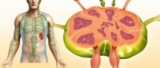
Location of the spleen relative to other organs
The spleen is an unpaired parenchymal organ, which is usually located in the left hypochondrium. It is covered on all sides by peritoneum and consists of several parts:
- upper and lower poles;
- diaphragmatic and visceral surfaces;
- gate (the splenic artery enters them, and the vein of the same name and nerves exit).
The outer, or diaphragmatic, surface of the organ is tightly adjacent to the costal section of the large muscle - the diaphragm. The projection of the spleen normally falls on the 9th-10th rib along the mid-axillary line and does not extend beyond the anterior lateral part of the body.
The lower pole of the organ is located at a distance of approximately 5-6 cm from the spinal column, which corresponds to the level of 10-11 thoracic vertebrae. The upper end of the spleen is in contact with the stomach and diaphragm, the lower end is in contact with the splenic angle of the colon. The tail of the pancreas also approaches the gate of the organ.
A non-enlarged parenchymal organ is not determined during palpation, and its length and width are calculated by percussion according to Kurlov. The size of the spleen is best determined by ultrasound. The technique makes it possible to measure its length and width.
Reasons for the increase
Possible causes of pathology
An enlarged spleen can occur due to various reasons. This pathology is called splenomegaly. The change in the size of the spleen is affected by stagnation of venous blood. Due to impaired blood flow, vascular tissue and the number of red blood cells grow, which leads to the development of Banti disease.
Congestion can occur when pressure in large veins increases due to a blood clot or severe heart failure.
The main reasons for an enlarged spleen are:
- Bacterial and viral infections
- Parasitic infections
- Neoplasms
- Liver diseases
- Blood diseases
- Hemolytic anemia
- Presence of cysts
In many cases, splenomegaly occurs as a result of various infections (hepatitis, rubella, measles, mononucleosis, etc.). The spleen is affected when helminths and arthropods enter the body.
The cause of an increase in the size of the spleen can be autoimmune diseases such as lupus erythematosus, rheumatoid arthritis, and periarteritis.
In case of damage to the spleen and the resulting tumors, ulcers, cysts or infarctions also affect the functioning of this organ. Formed elements in blood diseases are destroyed, and this entails an enlargement of the spleen. This is usually observed with hemolytic anemia, congenital spherocytosis, neutropenia, thrombocythemia, etc.
Symptoms
Signs of an enlarged spleen
With an enlarged spleen, no special signs are observed. Many may not know about this, and then discover the pathology during a medical examination.
Most often, clinical manifestations are observed with a significant size of the spleen, due to which surrounding organs are compressed.