Blockage of the tear duct, which occurs as a result of abnormal development of eye structures, inflammation, traumatic injury or other reasons, can lead to the most serious consequences. The OkoMed clinic offers adults and children treatment services for lacrimal duct obstruction .
The principle of operation of the tear duct
A tear is a biological fluid that performs important functions in the human body. It moistens the eye, protects it from dryness and saturates it with nutrients. In addition, tears have a disinfecting function and protect the mucous membrane from the effects of pathogenic microorganisms. Tear fluid is removed from the eye through the lacrimal ducts, and during the intrauterine development of the child they remain covered with a gelatinous film. It helps prevent amniotic fluid from entering the child’s body through the respiratory system.
After the baby is born, the gelatin film breaks and the tear duct opens. However, in some situations this does not happen and the child is diagnosed with congenital obstruction of the lacrimal duct. This pathology can be detected in older children and even adults.
Drops and massage
We saw an ophthalmologist only a month later, after a visit to a neonatologist. The doctor finally made a diagnosis and prescribed 2 more types of drops and massage of the tear duct. A follow-up visit was scheduled after 4 weeks.
My daughter’s drops didn’t work; they only made her eyes even more inflamed and festered. The only thing that really worked was washing with chamomile decoction, which, contrary to fears, did not cause dryness or irritation.
They didn’t really show me exactly how to do the massage, since the ophthalmologist refused to touch the child, and, as it later turned out, I understood her verbal explanations in my own way. In addition, to achieve a positive result, the procedure must be repeated 6 times a day, which they also forgot to tell me about.
In the end, of course, nothing changed significantly in a month. We cured the infection, but the eyes continued to water, which means that new inflammation was only a matter of time. The second time we were lucky to get to another specialist who approached the consultation much more responsibly. My daughter was prescribed more drops, and I finally received detailed instructions on how to do a massage. The next visit was due at 3 months of age.
I honestly tried to instill, rinse and massage with the regularity that was prescribed. But the problem was that the older Lisa became, the more negatively she perceived all these manipulations. At some point, I realized that I simply couldn’t cope with it alone. My daughter turned her head, grabbed my hands, and squirmed. She was not in pain, she just took any attempts to wash her eyes, give her a massage, clean her nose or ears with hostility, and began to scream and struggle. Now all this had to be done with 4 hands, and, as a result, there was simply no talk of any 6 times a day.
Causes of obstruction
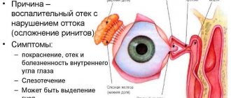
In adults, the main cause of dacryocystitis is narrowing and closure of the nasolacrimal duct, which causes impaired fluid circulation. The consequence of this pathological condition is stagnation of tear secretion, which provokes the active development of pathogenic microorganisms.
Experts identify the following reasons that can provoke the acquired form of pathology:
- Blockage by foreign objects. Dacryocystitis can occur when various chemicals, dust and cosmetics penetrate the tear ducts.
- Neoplasms of various types. The risk of problems with tear drainage increases with tumors in the nasal cavity, bone walls and lacrimal sac. Contact of tear fluid with the tumor causes its irritation, which aggravates the course of the pathology.
- Injuries and damage to the lacrimal ducts. With fractures of the bone walls of the orbit and nose, damage to the lacrimal ducts is often observed. Often, the probing of the nasolacrimal duct or its rinsing becomes a provoking factor for injury.
- Infectious lesions of the organs of vision. The risk of formation of adhesions in the nasolacrimal duct increases if the patient has pathological conditions such as rhinitis and chronic conjunctivitis.
Often the cause of damage to the lacrimal canal is a genetic predisposition to its occurrence. Constant relapses are observed in those people who have abnormalities in the structure of the eyelids and skull. The development of pathology is also possible with cleft palate, Down syndrome and deviations in the structure of the external nose.
How can you clarify the diagnosis?
Mothers often confuse symptoms of stenosis with signs of conjunctivitis and begin to independently treat the pathology at home. In this case, rinsing, compresses, and eye drops will not give the desired result. To understand the nature of the problem and eliminate it, an accurate diagnosis is necessary, which will be carried out by an ophthalmologist.
Diagnostic methods
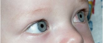
First, the specialist will examine and feel the baby’s eyes. This will allow you to determine whether there are protrusions in the corners of the eyes or whether there is tearing. By pressing on the lacrimal sac, the doctor assesses the nature of the released fluid. The ophthalmologist is also interested in:
- skin on the eyelids (for swelling and redness);
- eyelashes – uniformity and growth rate;
- condition of lacrimal openings.
Research methods conducted by the ophthalmologist himself will help make a final diagnosis.
Tests for dacryocystitis
| Name | What gives | Peculiarities |
| Kanaltsevaya | The condition of the lacrimal sac, tubules and points is assessed | The eyes are instilled with 2% collargol and checked after 5 minutes: • if the color has disappeared, there is no pathology; • the pigment dissolved within 10 minutes - the outflow of fluid was slowed down; • paint lingers for a long time in the conjunctival cavity - a clear sign of blockage. |
| Nasal | The entire system is checked for tear patency | The same product is dripped into the eyes as in the previous test. Turundas are rolled out of cotton wool and inserted into the sinuses, deepening them by 2 cm. Diagnosis is carried out according to the same principle as with the canalicular test: it is checked how many minutes it takes for the cotton swab to become colored. The absence of pigment is evidence of complete obstruction. |
In addition to the samples indicated in the table, an extensive laboratory blood test will be required. Eye discharge is also tested to detect infection.
Diagnostic method
Additional methods will help confirm or refute the suspected diagnosis:
- endoscopy of the nasal cavity;
- probing of the lacrimal ducts;
- flushing the ducts.
These methods have a dual purpose - they not only clarify the diagnosis, but are also a therapeutic measure.
In addition to an ophthalmologist, you will have to undergo examination by a pediatrician, ENT doctor, immunologist, infectious disease specialist and allergist to rule out the presence of ARVI, negative reactions of the body to irritants and a number of other factors. You may need to consult a surgeon.
Risk factors
There are some factors whose impact on the body can contribute to the development of the disease:
- disruption of metabolic processes in the body;
- diabetes;
- chronic eye inflammation;
- glaucoma;
- history of treatment of cancer;
- contact with irritants and the development of allergic reactions;
- decrease in the body's defenses.
Contact with various chemicals that affect the organs of vision can lead to dacryocystitis. In addition, atherosclerosis, tuberculosis and the pathological process in the lacrimal gland and its sac are considered provoking factors. Dacryocystitis is often observed in older women due to age-related changes.
Symptoms
With the disease, patients notice the appearance of:
- discomfort in the eye area;
- swelling of the lacrimal sac;
- increased lacrimation;
- mucous discharge mixed with purulent exudate;
- hyperemia and swelling of the conjunctiva;
- reduction in the size of the palpebral fissure;
- severe intoxication of the body.
A dense tumor is detected in the area of the lacrimal sac, which after some time begins to soften and the swelling disappears. During this period, an abscess develops, and it may open on its own, therefore, due to the removal of purulent exudate from it, tissue swelling decreases.
When the pathology passes into the chronic stage, the patient does not experience pain, but suffers from increased lacrimation. In addition, the edema in the area of the lacrimal canal transforms into a tumor, and when pressure is applied to it, yellow-green pus begins to be released from the lacrimal canaliculi.
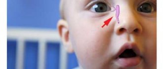
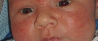
The first signs of a problem can be noticed almost immediately after birth
I noticed that the baby’s left eye began to leak while still in the maternity hospital on about the 3rd day. The neonatologist decided that the reason was that skin particles had gotten there. Just at this time, my daughter’s dry skin began to peel off, the top layer of which should have completely come off, so the theory could very well turn out to be correct. We were advised to wash our eyes with boiled water more often and after a couple of days we were sent home.
But ordinary rinsing did not have any effect, and when the doctor came to us after discharge, both eyes began to water and even fester. We were prescribed drops and a decoction of chamomile or a weak solution of furatsilin to clean the eyes, since they were supposedly infected in the maternity hospital. A week later, despite following all the recommendations, things did not get better, rather, quite the opposite, and by the time the doctor visited us again, the eyes were already quite festered.
As a result, Lisa was prescribed two more types of eye drops, one of which was an antibiotic. The situation improved a little, but my eyes continued to water. The suspicion that infection is not a cause, but a consequence, became increasingly substantiated.
Diagnosis of the disease
To make an accurate diagnosis, a visual examination of the patient is performed and instrumental techniques are used. A general blood and urine test is prescribed, after which a smear is made for bacteriological examination. Using this procedure, it is possible to identify the pathogenic culture that provoked the inflammatory process and select the most effective antibacterial drugs.
Some specific methods may be used to make a diagnosis:
- computed tomography of the naso-orbital zone;
- dacryocystography;
- probing of the lacrimal ducts.
Rhinoscopy is considered a mandatory diagnostic method, with the help of which it is possible to exclude pathologies caused by congenital anomalies of the structure of the nose. Medical practice shows that such disorders can provoke dacryocystitis, and surgical treatment is required to eliminate the problem.
A tubular test is required, during which a dye solution is instilled into the eyes. After the procedure, the specialist monitors the condition of the eyeball to determine whether this substance goes into the tubules.
Preparing the baby for probing
The operation is performed by a highly qualified pediatric ophthalmologist in a specially equipped eye office or clinic.
5 stages of preparation for the procedure:
- The final diagnosis should be confirmed by consultation with an otolaryngologist to ensure that there is no congenital curvature of the nasal septum.
- A complete blood test is taken to check clotting.
- A few hours before the operation, it is not allowed to feed the small patient to prevent the possibility of regurgitation during the procedure.
- Before the manipulation itself, the baby must be swaddled tightly to eliminate the possibility of chaotic movements of the arms and legs.
- It is better for the child to be well-rested and in a good mood.
Treatment of tear duct obstruction
With an uncomplicated course of the pathology, the prognosis is quite favorable. The choice of one method or another is determined by the form of the disease and the reasons for its development. Treatment of the pathology includes restoration of normal patency of the nasolacrimal duct and the use of anti-inflammatory drugs.
Conservative therapy
In most children, obstruction of the lacrimal duct disappears without treatment during the first months of his life. If this does not happen, then a special massage is prescribed. The use of drops containing antibiotics helps to cope with infections.
Conservative therapy is indicated for people whose pathology is identified at an early stage. The tubules are massaged and drops with an antibacterial effect are selected. If there is no positive effect, mechanical cleaning of the canals is carried out using a probe.
Conservative treatment includes prescribing:
- Tetracycline;
- Cefuroxime;
- Doxycycline;
- Chloramphenicol.
When an abscess develops, physiotherapy is usually prescribed, but it is best to perform an operation to open it.
Surgical methods
A procedure such as probing is performed on adult patients using local anesthesia. The specialist inserts a probe into the tear duct, after which it expands and removes the plugs. Usually it is possible to cope with the pathology in just one procedure, but repeated intervention is often required.
The operation is used to restore damaged tear ducts. Dacryocystorhinostomy is a type of surgical therapy, during which a new passage is formed to remove tears out.
Balloon dacryocystoplasty is considered the safest method of surgical treatment, which can be done even in childhood. When it is carried out, a special conductor is inserted into the nasolacrimal canal, which has a balloon filled with liquid. Under a certain pressure, it causes the ducts to expand, and thereby normalizes the movement of tear fluid.
How to treat dacryocystitis in children
Dacryocystitis of newborns involves three treatment options:
- conservative methods;
- wait-and-see tactics;
- surgical intervention.
Your doctor will determine which treatment method is right for you when examining your newborn. Do not self-medicate or use unconventional folk methods. A newborn is not a field for experimentation.
Conservative methods of treating dacryocystitis include medications and massage. Combining these two methods can significantly speed up the healing process and alleviate the condition of the newborn baby.
Drug treatment
Obstruction of the nasolacrimal duct in children is treated mainly with drops and ointments. The choice of antibacterial agent should be based on seeding and seeded microflora. Drops are instilled during the day and after a massage, and ointments are placed behind the lower eyelid at night. The dosage and method of administration are prescribed by the doctor.
Drops and ointments for dacryocystitis for the treatment of newborns:
- "Albucid".
- Vigamox.
- Tobrex is often prescribed to infants.
- "Levomycetin".
- Gentamicin ointment.
- Dexamethasone ointment.
- Oftaquix.
- A solution of furatsilin or chlorhexidine for washing and wiping the eyes.
Before use, the drops must be warmed to body temperature in the palm of your hand or in a water bath. Since opened medications must be stored in the refrigerator, it will be very unpleasant for the baby to drop cold medications into the eye.
Watch an interesting video with advice from a pediatrician about treating the disease:
Massage
How to pierce the tear duct yourself without surgery? The main method of treating dacryocystitis in newborns is a special massage. The movements resemble pressure from the corner of the eye to the tip of the nose along the nasal septum. This physically pushes out any blockages and helps the tubules free up.
Massage technique for newborns with dacryocystitis:
- First of all, you need to wash your hands, remove all jewelry, and trim your nails so as not to injure the newborn or cause an infection.
- If purulent discharge is present, first, using a movement from below, it is necessary to squeeze out the purulent contents. Wipe your eye with a cotton pad or gauze soaked in an antiseptic solution.
- Then instill the antibiotics in drops and now push the drops from top to bottom through the tubules into the lacrimal sac and beyond. Drops must be instilled several times.
- Repeat these movements ten times, two to three times a day. At night, place ointment behind the lower eyelid.
What the country’s most famous pediatrician, Dr. Komarovsky, says about massage for inflammation of the tear duct in children, can be seen here: