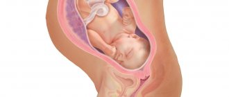
Most babies are born with a head. That is, the baby is placed upside down in the uterine cavity. This position of the baby is called cephalic presentation.
If a pregnant woman is not told before 36 weeks that her baby is in a breech position, there is no need to worry. After all, the baby is constantly changing position; some children at 32 weeks may be feet down.
After the 36th week, the amount of amniotic fluid becomes less, and it is more difficult for the fetus to change its position.
Fetal presentation is the relationship of the large part of the fetus to the inlet of the pelvis.
If the head is located above the entrance to the woman’s pelvis, the presentation is cephalic , if the pelvic end is pelvic.
The presenting part is the lowest located part of the fetus, which is the first to pass through the birth canal.
What does placenta previa mean?
The placenta is a temporary organ and appears only during pregnancy. With the help of the placenta, the mother and fetus communicate, the child receives nutrients through its blood vessels and gas exchange occurs. If the pregnancy proceeds normally, the placenta is located in the area of the fundus of the uterus or in the area of its walls, usually along the back wall, moving to the sides (in these places the blood supply to the muscle layer is more intense).
Placenta previa is said to be present when the latter is located incorrectly in the uterus, in the area of the lower segment. In fact, placenta previa is when it blocks the internal os, partially or completely, and is located below the presenting part of the baby, thus blocking the path for birth.
“Boot to the world” and more
Breech presentation of the fetus indicates that the pelvic end is located at the entrance to the small pelvis.
The prevalence of breech presentation is about 5%.
Breech presentation is more common in multiparous women.
As pregnancy progresses, the frequency of breech presentations decreases.
The risk of maternal mortality with breech presentation is about 2%, it is associated with traumatic injuries, bleeding during childbirth, infection of the membranes and the fetus.
Childbirth with a breech presentation can have a normal course, but complications that are dangerous for the mother and fetus cannot be ruled out. Therefore, such births will also differ in the nature of the interventions, since they can lead to complications in the form of injury to the newborn, including death. Such births are called pathological, and the pregnant woman and child are classified as a high-risk group.
What is breech presentation like?
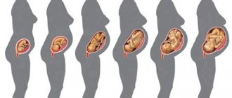
- breech presentation (flexion):
- purely gluteal (incomplete). Classic version: the child lies with his butt down, his body is folded in half, his legs are straightened, his feet are at face level.
- mixed gluteals (full). In this case, the child squats.
- foot presentation (extensor):
- full;
- incomplete;
- knee
Only 3% of children are born in a breech or leg presentation (the buttocks or legs emerge first from the birth canal).
Pure breech presentation (in first-time mothers) is more common, mixed breech presentation is less common, and even less common is foot and knee presentation.
If a woman is diagnosed with breech presentation at a later stage, she must fulfill certain conditions:
- exclusion of sudden movements, bending forward, sleeping on the back;
- It is prohibited to wear a bandage.
Types of choreon presentation
There are several classifications of the described obstetric pathology. The following is generally accepted:
Separately, it is worth highlighting low placentation or low placenta previa during pregnancy.
Low placentation is the localization of the placenta at a level of 5 or less centimeters from the internal os in the third trimester and at a level of 7 or less centimeters from the internal os in pregnancy up to 26 weeks.
A low location of the placenta is the most favorable option; bleeding during gestation and childbirth rarely occurs, and the placenta itself is prone to so-called migration, that is, an increase in the distance between it and the internal os. This is due to the stretching of the lower segment at the end of the second and third trimesters and the growth of the placenta in the direction that is better supplied with blood, that is, to the uterine fundus.
In addition, the presenting vessels are identified. In this case, the vessel/vessels are located in shells, which are located in the area of the internal pharynx. This complication poses a threat to the fetus if the integrity of the vessel is damaged.
Classification
According to the domestic classification, the following types of breech presentations are distinguished:
- Gluteal or flexor
- purely gluteal - when the buttocks are adjacent to the entrance, and the legs are bent at the hip joints, but extended along the body of the fetus and press the arms to the chest, and the head is also pressed to the chest;
- mixed gluteal – when the buttocks and foot (one or both) are adjacent to the entrance;
- Leg or extensor
- incomplete leg - when only one leg is adjacent to the entrance (and nothing else);
- full leg – respectively, both legs are adjacent;
- knee - the fetus seems to be on its knees, it is quite rare, and during the birth process it turns into a leg position.
Most often, pure breech presentations are observed (up to 68% of all breech presentations), mixed breech presentation in 25%, and foot presentation in 13%. During childbirth, it is possible to transition from one type of breech presentation to another. Complete leg is diagnosed in 5 - 10%, and incomplete leg is observed in 25 - 35% of births.
Expectant mothers should not immediately become upset because the baby is lying incorrectly. A lot of fetuses that are presented at the pelvic end by the end of pregnancy turn over and are presented at the head.
Such spontaneous rotation is more often observed with the presentation of the buttocks, and in multiparous women this happens 2 times more often than in first-born women. And, what’s good is that if the child turned over on his own, then his reverse “somersault” is unlikely.
Provoking factors
The reasons that cause placenta previa can be associated both with the condition of the mother’s body and with the characteristics of the fetal egg. The main reason for the development of complications is degenerative processes in the uterine mucosa. Then the fertilized egg is not able to penetrate (implant) into the endometrium of the fundus and/or body of the uterus, which forces it to descend lower. Predisposing factors:
- chronic inflammation of the uterus;
- numerous births;
- abortions and uterine curettage;
- childbirth and abortion complicated by purulent-septic diseases;
- uterine tumors;
- scars on the uterus (operative delivery, removal of fibroids);
- congenital anomalies of the uterus;
- internal endometriosis;
- sexual infantilism;
- smoking;
- drug use;
- first birth at age 30 or more;
- impaired hormonal function of the ovaries;
- multiple pregnancy.
Chronic endometritis, numerous intrauterine manipulations (curettage and abortion), myomatous nodes lead to the formation of an incomplete second phase of the endometrium, in which it prepares for implantation of a fertilized egg. Therefore, when forming the chorion, she looks for the most favorable place, which is well supplied with blood and optimal for placentation.
Symptoms of placenta previa
Symptoms of placenta previa are explained by its incorrect localization. The pathognomic sign of this obstetric complication is external bleeding. Bleeding from the uterus can occur at any stage of pregnancy, but more often in the last weeks of gestation. This has two reasons.
Typically, bleeding always begins suddenly, often against the background of absolute rest, for example, in sleep. It is impossible to predict when bleeding will occur and how intense it will be.
Of course, the percentage of profuse bleeding with central presentation is much greater than with incomplete presentation, but this is not necessary. The longer the gestational age, the greater the chance of bleeding.
- For example, marginal placenta previa may not manifest itself at all at 20 weeks, and bleeding will occur (but not necessarily) only during childbirth.
- Low placentation most often occurs without clinical symptoms, pregnancy and childbirth proceed without any special features.
One of the typical characteristics of bleeding during presentation is its recurrence. That is, every pregnant woman should know about this and always be on guard.
- The volume of bleeding varies: from intense to insignificant.
- The color of the blood released is always scarlet, and the bleeding is painless.
Any minor factor can provoke bleeding:
- straining during bowel movements or urination
- cough
- sexual intercourse or vaginal examination
Another difference between placenta previa is the woman’s progressive anemia (see low hemoglobin during pregnancy). The volume of blood lost almost always does not correspond to the degree of anemia, which is much higher. During repeated bleeding, the blood does not have time to regenerate, its volume remains low, which leads to reduced blood pressure, the development of disseminated intravascular coagulation syndrome or hypovolemic shock.
Due to the incorrect location of the placenta, progressive anemia and reduced volume of circulating blood, fetoplacental insufficiency develops, which leads to intrauterine growth retardation and the occurrence of intrauterine hypoxia.
Example from practice: A 35-year-old woman was observed in a antenatal clinic; she was pregnant for the second time, and was wanted. At the first ultrasound at 12 weeks, she was diagnosed with central placenta previa. An explanatory conversation was held with the pregnant woman, and appropriate recommendations were given, but my colleague and I observed with fear and expectation of bleeding. During the entire period of pregnancy, she experienced bleeding only once, at 28–29 weeks, and even then, it was not bleeding, but minor bloody discharge. Almost the entire pregnancy, the woman was on sick leave; she was hospitalized in the pathology ward at dangerous times and during the period of bleeding. The woman safely reached term and at 36 weeks was sent to the maternity ward, where she successfully prepared for the upcoming planned caesarean section. But, as often happens, on a holiday she started bleeding. Therefore, an operating team was immediately convened. The baby was born wonderful, even without signs of malnutrition (in children). The afterbirth was separated without problems, the uterus contracted well. The postoperative period also proceeded smoothly. Of course, everyone breathed a sigh of relief that such a huge burden had been lifted from their shoulders. But this case is rather atypical for central presentation, and the woman, one might say, was lucky that everything ended with little bloodshed.
Course of pregnancy
The final diagnosis of breech presentation is made at 36 weeks, when the fetus is firmly in position in the uterus, although spontaneous rotation is not excluded. Pregnancy with a breech presentation of the fetus is much more likely to have complications than with a cephalic presentation. The main complications are:
- threat of miscarriage or premature birth;
- gestosis;
- placental insufficiency.
All of these complications lead to oxygen starvation of the fetus, and, accordingly, to its developmental delay (hypotrophy and low weight), abnormal amount of amniotic fluid (low or polyhydramnios), and entanglement of the umbilical cord. In addition, breech presentation is often accompanied by placenta previa, unstable fetal position and prenatal rupture of water.
Also, such presentation affects the development of the fetus and the functions of the fetoplacental system:
- Maturation of the medulla oblongata
By 33–36 weeks, the maturation of the medulla oblongata begins to slow down, which is manifested by pericellular and perivascular edema of the brain, which leads to “swelling” and impaired blood circulation in the brain, and, consequently, to a disorder of its functions.
The function of the adrenal glands, as well as the hypothalamic-pituitary system, is depleted, which significantly reduces the adaptive and protective reactions of the fetus during childbirth and after.
- Sex gonads (testes and ovaries)
There is poor circulation and tissue swelling, mature cells of the sex gonads partially die, which subsequently affects reproductive function (hypogonadism, oligo- and azoospermia) and leads to infertility.
- Congenital malformations
When presented with the pelvic end, congenital defects occur 3 times more often, in contrast to cephalic presentation. Primarily, defects of the central nervous system and heart, as well as anomalies of the digestive tract and musculoskeletal system.
- Disturbance of uteroplacental blood flow
Leads to fetal hypoxia, increased heart rate and decreased motor activity.
How to diagnose?
Placenta previa is a hidden and dangerous pathology. If the pregnant woman has not yet had bleeding, then presentation can be suspected, but the diagnosis can only be confirmed using additional examination methods.
A carefully collected anamnesis (in the past there were complicated childbirths and/or the postpartum period, numerous abortions, diseases of the uterus and appendages, operations on the uterus, etc.), the course of the current pregnancy (often complicated by the threat of miscarriage) and external obstetric data helps to suggest a placenta previa. research.
During an external examination, the height of the uterine fundus is measured, which is greater than the expected gestational age, as well as abnormal position of the fetus or breech presentation. Palpation of the presenting part does not give clear sensations, as it is hidden under the placenta.
If a pregnant woman complains of bleeding, she is hospitalized in a hospital to exclude or confirm the diagnosis of such a pathology, where, if possible, an ultrasound is performed, preferably with a vaginal sensor. A speculum examination is carried out to determine the source of bloody discharge (from the cervix or varicose veins of the vagina).
The main condition that must be observed when examining with mirrors: the examination is carried out against the backdrop of a deployed operating room and always with heated mirrors, so that in case of increased bleeding, the operation can be started without delay.
Ultrasound remains the safest and most accurate method for determining this pathology. In 98% of cases, the diagnosis is confirmed; false positive results are observed when the bladder is overly full, so when examined with an ultrasound probe, the bladder should be moderately full.
Ultrasound examination allows not only to determine the presentation of the choreon, but also to determine its type, as well as the area of the placenta. The timing of ultrasound examinations during the entire period of gestation is somewhat different from the timing of normal pregnancy and corresponds to 16, 24 - 26 and 34 - 36 weeks.
Million dollar tips on how to get your baby to roll over
Attempts to turn the baby around are recommended from 32 to 37 weeks, then the amount of amniotic fluid decreases and the child becomes less active in his movements.
Exercises cannot be performed:
- immediately after eating;
- for hypertension;
- with hypotension;
- with polyhydramnios;
- with gestosis;
- with placenta previa;
- if pain occurs during exercise;
- with too high fetal activity.
External obstetric rotation (Arkhangelsky method)
Performed by an experienced doctor at 36-37 (for first-time mothers) and 38 weeks (for multiparous women) of pregnancy under ultrasound control and monitoring the condition of the fetus using CTG. The method gives the baby the opportunity to roll over on his own and take his physiological position. The fetus makes a somersault with the help of the doctor's hands. The method can lead to premature birth, so the woman should be prepared for such a development. If you can help the baby turn over, the birth takes place naturally.
Internal turn
It is carried out only during childbirth. Due to the large number of complications, it is rarely used.
One of the most effective exercises is “Inversion”
To prevent fetal malposition, American midwife Gail Tully came up with an exercise that relaxes the uterosacral ligaments and creates more space in the lower segment of the uterus, as a result the baby is able to change its position.
The exercise is performed every day, starting from the 35th week of pregnancy. It is recommended to perform it during periods when the baby is active.
The duration of the exercise is about 10-15 minutes.
There are several variations of poses for this exercise.
Main pose
Technique:
- you need to kneel at the edge of the bed or sofa;
- Gently lower your hands to the floor, with your forearms touching the floor and connect your fingers together;
- the head should hang freely in the air without touching the floor;
- you are allowed to slightly swing your hips during the exercise;
- There is no need to bend the spine too much in the lumbar region, this will help to better relax the ligaments.
Tilt on a hard surface
Performed immediately after the main pose daily for 20 minutes 3 times a day.
You need a hard surface (like an ironing board), which is placed at an angle of 45 degrees.
The woman lies down on it so that her head is below and her pelvis is above her head.
The baby will get more space with this exercise.
Knee-elbow pose
The arms and legs are spread wide apart, the head rests on the hands. Perform up to three times a day for 10-15 minutes.
Raising the pelvis
While lying on your back, you need to raise your pelvis. This can also be done 3 times a day for 15 minutes.
The last two poses are alternatives to the second pose. You can change them after completing the first main exercise.
Turns
A pregnant woman turns over onto her right and left side while lying on the couch for 10 minutes on each side, repeating the procedure three times.
The course lasts 10 days, 3 times a day.
Sifting method using a Rebozo scarf
There is a special Mexican Rebozo scarf, which is spread under the back of a lying woman. It covers the pelvic bones, relieves tension from the spine, and reduces lower back pain.
To turn the baby over, a pregnant woman kneels and rests her hands on any support. At this time, the specialist hugs her stomach with a scarf, massaging it with trembling light movements.
Webster's method
Gluteal technique of American doctor Larry Webster (intrauterine pressure method). It was designed to restore normal pelvic balance and function of the pelvic organs.
Performed from 36 weeks, it helps to relax the uterus and ligaments, allows you to relax the muscles of the anterior abdominal wall, which facilitates the natural turning of the baby. Helps cope with back pain.
Thanks to this technique, the sacrum and pelvic bones are in a balanced and correct state, and the uterine ligaments are relaxed: the baby has more room to move.
This procedure is carried out under the supervision of a specialist.
Hot-cold method
A cold compress is placed on the upper abdomen, and a warm compress is placed on the lower abdomen. This may encourage the baby to move away from the cold towards the warm, turning over into the correct position.
Swimming
Being in the water and doing simple exercises can help your baby roll into the desired position.
Treatment with music
The headphones are applied to the lower abdomen, the child should turn towards the sound source.
Conversations with father
Communication between dad and baby often helps the child obey his father and roll over on his own.
Breath
Lying on your back, legs bent at the knees. A pregnant woman inhales through her nose, while her stomach inflates, and when she exhales, she deflates her stomach.
Visualization
The woman imagines how her baby turns over in his stomach and takes the correct position. You can look at the pictures.
Chinese medicine method (moxibustion)
A smoldering stick of dried wormwood is located at a distance of 6 mm from the little toe of the pregnant woman (point BL 67), which is located near the outer corner of the fifth nail on the baby’s foot. The method works by increasing the baby’s physical activity. Performed by an acupuncturist. This method is not used in Russia (Bali).
How pregnant women are managed and delivered
If placenta previa is confirmed, treatment depends on many circumstances. First of all, the period of pregnancy when bleeding occurred, its intensity, the amount of blood loss, the general condition of the pregnant woman and the readiness of the birth canal are taken into account.
If chorionic presentation was established in the first 16 weeks, there is no bleeding and the woman’s general condition does not suffer, then she is treated on an outpatient basis, having previously explained the risks and given the necessary recommendations (sexual rest, limitation of physical activity, prohibition of taking baths, visiting baths and saunas).
Upon reaching 24 weeks, the pregnant woman is hospitalized in a hospital, where preventive therapy is carried out. Also, all women with bleeding are subject to hospitalization, regardless of its intensity and stage of pregnancy. Treatment of the described obstetric pathology includes:
- medical and protective regime;
- treatment of fetoplacental insufficiency;
- anemia therapy;
- tocolysis (prevention of uterine contractions).
The protective treatment regime includes:
- prescription of sedatives (tincture of peony, motherwort or valerian)
- maximum restriction of physical activity (bed rest).
- Therapy of fetoplacental insufficiency prevents fetal development delay and consists of prescribing: antiplatelet agents to improve the rheological qualities of the blood (trental, chimes)
- vitamins (folic acid, vitamins C and E)
- actovegin, cocarboxylase
- Essentiale-Forte and other metabolic drugs
- It is mandatory to take iron supplements to increase hemoglobin (sorbifer-durule c, tardiferon and others).
Tocolytic therapy is carried out not only in the case of a threatened miscarriage or threatening premature birth, but also for the purpose of prevention, the following are indicated:
- antispasmodics (no-spa, magne-B6, magnesium sulfate)
- tocolytics (ginipral, partusisten), which are administered intravenously.
- in the case of threatening or beginning premature labor, prevention of respiratory disorders with corticosteroids and (dexamethasone, hydrocortisone) is mandatory for a duration of 2–3 days.
If bleeding occurs, the intensity of which threatens the woman’s life, regardless of the gestational age and the condition of the fetus (dead or nonviable), abdominal delivery is performed.
What to do and how to deliver a child with chorionic presentation? Doctors ask this question when they reach 37–38 weeks. If there is a lateral or marginal presentation and there is no bleeding, then in this case the tactics are expectant (the beginning of spontaneous labor). When the cervix is dilated by 3 centimeters, an amniotomy is performed for prophylactic purposes.
If bleeding occurs before the onset of regular contractions and there is a soft and distensible cervix, an amniotomy is also performed. In this case, the baby’s head lowers and is pressed against the entrance to the pelvis, and, accordingly, presses the detached lobules of the placenta, which causes the bleeding to stop. If the amniotomy has no effect, the woman is delivered abdominally.
Caesarean section is routinely performed for those pregnant women who have been diagnosed with complete presentation, or in the presence of incomplete presentation and concomitant pathology (improper position of the fetus, pelvic end presentation, age, uterine scar, etc.). Moreover, the surgical technique depends on which wall the placenta is located on. If the placenta is localized along the anterior wall, a corporal cesarean section is performed.
Text of the book “Obstetrics and Gynecology: Lecture Notes”
Physical examination
A physical examination is carried out taking into account the history and complaints of the pregnant woman.
At the same time, attention is paid to those organs whose diseases were observed previously. During the first stage of labor, the examination is carried out between contractions. General inspection
Basic physiological indicators.
The pulse rate is measured, blood pressure is measured in pauses between contractions. If necessary, the measurement is carried out several times. A sign of chorioamnionitis may be an increase in body temperature, especially after the rupture of amniotic fluid. Tachycardia and tachypnosis during labor in the absence of changes in other physiological parameters are normal.
An ophthalmoscopy is necessary to exclude retinal hemorrhage, vasospasm, or retinal edema, which may be present in diabetes mellitus and arterial hypertension. Pale conjunctivae or nail beds may be a sign of anemia. Swelling of the face, hands and feet is observed with preeclampsia. Palpation of the thyroid gland is mandatory.
A rare but serious complication during childbirth - venous congestion is manifested by swelling of the neck veins and requires mandatory treatment. If a woman has a history of bronchial asthma, auscultation of the lungs is performed to detect shortness of breath and wheezing and auscultation of the heart, paying attention to the presence of systolic murmur. It must be remembered that mesosystolic murmur is observed during pregnancy normally.
The abdomen is palpated to exclude pain and the presence of space-occupying formations. Pain on palpation of the epigastric region may be a sign of preeclampsia. During full-term pregnancy, palpation of the abdomen is difficult.
During full-term pregnancy, minor swelling of the legs is normal. A neurological examination is carried out if severe swelling of the legs or hands is detected (signs of preeclampsia). Increased tendon reflexes and clonus indicate increased seizure readiness.
External obstetric examination
Dimensions of the uterus.
By the end of the 1st obstetric month (4th week), the uterus reaches the size of a chicken egg. It is usually not possible to determine pregnancy with a vaginal examination. By the end of the 2nd month (8th week), the uterus increases to the size of a goose egg. By the end of the 3rd month (12th week), asymmetry of the uterus (Piskachek’s sign) is noted; it increases to the size of a man’s fist, its bottom reaches the upper edge of the symphysis. By the end of the 4th month (16th week), the uterine fundus is determined at the middle of the distance between the symphysis and the navel or 6 cm above the navel. By the end of the 5th month (20th week), the fundus of the uterus is located 11–12 cm above the womb or 4 cm below the navel. By the end of the 6th month (24th week), the fundus of the uterus is at the level of the navel or 22–24 cm above the womb. By the end of the 7th month (28th week), the uterine fundus is determined two transverse fingers above the navel or 25–28 cm above the womb. By the end of the 8th month (32nd week), the fundus of the uterus is located in the middle of the distance between the navel and the xiphoid process, 30–32 cm above the pubis. By the end of the 9th month (36th week), the fundus of the uterus reaches the xiphoid process and costal arc. By the end of the 10th month (40th week), the fundus of the uterus drops to the level of a 32-week pregnancy. By palpation of the uterus, the approximate size of the fetus and the amount of amniotic fluid are determined. It is also important to determine the thickness of the anterior abdominal wall of the woman in labor and the degree of insertion of the presenting part of the fetus into the pelvic area. It is necessary to exclude malformations of the uterus or fetus or multiple pregnancies if the size of the uterus exceeds the expected gestational age. For this purpose, an ultrasound is performed.
External obstetric examination includes four Leopold maneuvers.
First appointment
allows you to determine the height of the uterine fundus and that part of the fetus that is located in the uterine fundus. The head is more rounded and dense compared to the buttocks. The head moves, and the pelvic part moves only along with the fetal body.
Second appointment
serves to determine the position of the fetus and its type. Consists of palpation of the lateral surfaces of the uterus.
It allows you to determine on which side the small parts of the fetus are located (arms, legs), and on which side the back is located, as well as its movement, the tone of the uterus.
Third appointment
used to determine the presenting part and its relationship to the entrance to the pelvis. The head must be distinguished from the pelvic end of the fetus. It is round and dense. When the head moves, the symptom of balloting is noted. In case of breech presentation, a bulky part of the fetus with a softish consistency without clear contours is determined above the entrance to the pelvis, which does not give the symptom of balloting. By shifting the presenting part from side to side, its position is determined in relation to the entrance to the pelvis. If displacement is difficult, it means that it is fixed at the entrance to the pelvis.
Fourth technique
allows you to clarify the presentation of the fetus. To perform the maneuver, the obstetrician turns to face the woman in labor and palpates the presenting part with both hands. With an occipital presentation, the occipital curvature is determined on the same side as the small parts of the fetus, while the head is bent and the occiput is presented. With a facial presentation, the occipital curvature is determined on the opposite side of the small parts of the fetus, the head is extended.
The location of the fetus in the uterus.
According to the basic research methods, it is possible to easily determine the position of the fetus in the uterus, its position, position and type of fetus.
Fetal position
- this is the ratio of the longitudinal axis of the fetal body to the longitudinal axis of the mother’s body. The position of the fetus can be longitudinal (with pelvic or cephalic presentation), transverse and oblique, when the axes of the fetal and maternal bodies intersect. The articulation of the fetus is the relationship of the fetal limbs and head to its body. A favorable articulation is the flexion type, in which the fetus resembles an ovoid in appearance.
Fetal presentation.
This is the relationship of the large part of the fetus to the entrance to the pelvis.
The presenting part is the part of the fetal body that is located above the entrance to the pelvis. The fetal head, pelvis or shoulder may be present. The most common and physiological is considered to be cephalic presentation. When the fetal head is flexed, the presentation will be considered occipital. When the head is in an extension position, a frontal or facial presentation is formed. If the pelvic part of the fetus is located above the entrance to the pelvis, the presentation is called pelvic
. Breech presentation can be purely breech (the legs of the fetus are extended along the body, and the buttocks are facing the entrance to the pelvis), mixed breech (the buttocks and feet of the fetus are presented), complete leg (both legs are presented) and incomplete (one leg is presented). With foot presentation, a complication often occurs in the form of umbilical cord prolapse. In the transverse position, the fetal shoulder is located above the entrance to the pelvis. In a normal full-term pregnancy, it is very rare that several parts of the fetal body (head and small parts) may present simultaneously.
Fetal position
called the relationship of the fetal back to the left or right wall of the uterus. There are first (left) and second (right) positions of the fetus.
Type of fruit
– the ratio of its back to the anterior wall of the uterus. The first position is often combined with a front view, the second - with a rear view.
Auscultation
Fetal heart tests have recently increasingly been replaced by CTG. This method helps to more accurately record heart rate and heart rate variability (acceleration and deceleration).
Conducting a vaginal examination
It begins with inspection and palpation of the perineal and pelvic area. In the presence of bleeding from the vagina and premature discharge of amniotic fluid, a vaginal examination is carried out only after an ultrasound.
Examination of the perineum consists of identifying herpetic eruptions, varicose veins of the external genitalia, the presence of condylomas, and scars. In cases of suspected labia herpes, a thorough examination of the cervix and vagina is necessary. Also, during the examination, attention is paid to the integrity of the pelvic bones and amniotic sac, the opening and smoothing of the cervix, as well as the position of the presenting part.
Diagnosis of discharge of amniotic fluid
almost never in doubt, but if necessary, examine the cervix and vaginal vault in the speculum. When amniotic fluid ruptures, a vaginal examination may reveal the fetal buttocks, or the head or loops of the umbilical cord. In this case, amniotic fluid is present in the posterior vaginal fornix. If the fluid present in the posterior fornix contains amniotic fluid, then a microscopic examination of the dried smear reveals a fern phenomenon. Amniotic fluid turns the test strip dark blue if the result is positive, as it has an alkaline reaction. The test may be false positive if there is blood or urine in the posterior fornix. The possible admixture of meconium is also taken into account. Meconium is the primary fecal content of the fetal intestine, which increases in late pregnancy. The presence of meconium in the amniotic fluid is a sign of fetal hypoxia. The presence of blood in the amniotic fluid may be a sign of placental abruption. If premature birth occurs and chorioamnionitis is suspected, a culture of discharge from the posterior vaginal fornix is performed. In case of premature rupture of amniotic fluid, it is necessary to determine the degree of maturity of the fetal pulmonary system using a foam test.
Cervix
Degree of opening
The cervix is measured in centimeters: from 0 (the cervix is closed) to 10 cm (fully dilated).
Cervical smoothing
is one of the indicators of her maturity and readiness for childbirth. The size of the immature cervix is 3 cm (degree of effacement 0%). Smoothing occurs gradually and becomes maximum at the beginning of labor (100% degree of effacement). In primiparous women, the cervix first undergoes effacement and then dilatation. During repeated births, effacement and dilatation of the cervix occur almost simultaneously.
Palpation of the presenting part of the fetus
Fetal presentation
determined by palpation. With an occipital presentation, you can palpate the sutures and fontanelles on the fetal head, with a pelvic presentation, you can identify the buttocks and feet, with a facial presentation, you can palpate the front part of the fetal head, but ultrasound provides more accurate data on the presentation.
The degree of insertion of the presenting part into the pelvis.
In order to determine the position of the presenting part, the line that connects the woman’s ischial spines is taken as the starting point. If, during an occipital presentation, the fetal head has reached this line, it means that it has entered the small pelvis with biparietal size (degree of insertion “0”). If the presenting part is 1 cm above the ischial spines, the degree of its insertion is determined as “–1”, if 2 cm below the ischial spines - as “+2”. If the degree of insertion of the presenting part is more than “-3”, then this means its mobility above the entrance to the pelvis. If the degree of insertion is “+3”, then the presenting part is located at the bottom of the pelvis and is visible in the genital slit during pushing.
Fetal position
- this is the location of certain points of the presenting part of the fetus in relation to the anatomical structures of the pelvis. In the anterior position, the presenting part faces the pubic symphysis; in the posterior position, it faces the sacrum. Transverse (right or left) position - the presenting part faces the right or left wall of the pelvis. The position for occipital presentation can be determined by the point of intersection of the lambdoid and sagittal sutures. In case of pelvic presentation - along the sacrum of the fetus, in case of facial presentation - according to the location of the chin. In the anterior position of the occipital presentation, the back of the head is turned towards the pubic symphysis. In the right transverse position of the occipital presentation - to the right vaginal wall.
Examination of the pelvic bones.
The size and shape of the small pelvis are determined by the size of the large pelvis. A narrow pelvis has such features that when a full-term fetus passes through it, mechanical obstacles are formed. External measurement of the pelvis does not always make it possible to identify the shape and degree of narrowing of the pelvis. In some cases, the discrepancy between the sizes of the pelvis and the fetal head can only be determined during childbirth. The size of the pelvis is one of the three main factors that determine the physiological course of childbirth. Other important factors are the size of the fetus and normal labor.
Entrance to the pelvis
in front it is formed by the upper edge of the pubic articulation of the symphysis, in the back - by the apex of the promontorium, the lateral borders are by the arcuate lines of the ilium. The direct size of the small pelvis is determined by the size of the diagonal conjugate - the distance between the promontory and the lower edge of the pubic symphysis, which is normally 12 cm or more.
Pelvic cavity
in front it is formed by the posterior surface of the pubic symphysis, in the back by the anterior surface of the sacral vertebrae, and the lateral walls by the ischial bones. The transverse size of the pelvic cavity is normally more than 9 cm. This distance between the ischial spines is determined during a vaginal examination.
Exit from the pelvis
in front it is located in the area of the lower edge of the pubic arch, in the back - in the area of the apex of the coccyx, on the sides - between the ischial tuberosities. The transverse size of the small pelvis is the distance between the ischial tuberosities of at least 8 cm at normal sizes. You can indirectly judge the size of the outlet from the small pelvis by the size of the subpubic angle and the protrusion of the apex of the coccyx. An acute subpubic angle most often indicates a narrow pelvis. Usually there is a combined decrease in all sizes of the small pelvis.
Laboratory research
Mothers in labor undergo laboratory tests: a general blood and urine test and a serological test for syphilis, and if they belong to a high-risk group, for HBsAg. If there is a history of changes in glucose levels, then the blood glucose level is determined. If preeclampsia is suspected, platelet counts are calculated, liver and kidney functions, and serum uric acid levels are determined. If premature placental abruption is suspected, a coagulogram is examined. If there is a possibility of a cesarean section, or a history of bleeding in the afterbirth period, or multiple births, the pregnant woman’s serum is stored to quickly test for individual compatibility. If concomitant complications or diseases are identified in the anamnesis and during examination, additional laboratory tests are performed. If a history or physical examination reveals any concomitant diseases or complications, additional laboratory tests are prescribed.
Assessment of fetal condition during labor
It is carried out for the purpose of early diagnosis of intrauterine hypoxia and fetal death. For this purpose, a number of examinations are carried out: auscultation of the fetal heart at certain intervals, continuous CTG (direct or indirect), determination of the acid-base composition of blood obtained from the scalp.
Auscultation of the fetal heart in the first stage of labor is carried out every 15 minutes, and in the second stage of labor - every 5 minutes (or after each attempt).
Formulation of the conclusion and labor management plan.
Anamnesis collection, examination and laboratory data allow us to determine the prognosis and tactics of labor management.
Lecture No. 6. Physiological childbirth
Childbirth is a staged physiological process during which the expulsion of the fetus occurs, as well as the release of amniotic fluid, membranes and placenta through the natural birth canal.
Urgent
Childbirth is considered to be in the 37th–42nd week of pregnancy, and
premature
birth is considered to be birth before the 37th week of pregnancy.
Childbirth after the 42nd week of pregnancy is called late
.
Periods of labor
The disclosure period is the first period. It starts with the first contraction. They are frequent, intense, long-lasting. During contractions, the cervix softens and dilates. The first period is divided into two phases - the slow opening phase and the fast opening phrase. During the first phase, the cervix dilates up to 4 cm, during the second - from 4 to 10 cm. The transition from contractions to pushing and the discharge of amniotic fluid completes the first period.
The second period (expulsion) is characterized by the expulsion of the fetus. At this time, the cervix is fully dilated, contractions turn into pushing. The second period ends with the birth of a child.
Third period (successive). The beginning is the moment of birth of the child, the end is the separation of the placenta and the birth of the placenta.
Labor assessment criteria are based on cervical effacement and dilatation and advancement of the presenting part of the fetus. At the same time, frequent vaginal examinations should not be carried out in order to avoid infection of the fetus and not cause discomfort to the mother in labor.
First stage of labor
Its duration for first-time mothers is about 12 hours, for repeat births – about 7 hours.
Immediately after the onset of contractions, it is necessary to monitor the fetal heart rate. Fetal hypoxia (begun or threatening) may be indicated by a sudden increase in heart rate (more than 140 per minute) or a decrease (less than 120 per minute).
Amniotomy is performed for the following indications:
1) polyhydramnios, flat membranes, marginal placenta previa, premature placental abruption;
2) the need for direct access to the fetus for invasive procedures;
3) labor induction and labor stimulation.
Only amniotomy performed in the rapid dilatation phase has a birth-stimulating effect. Amniotomy performed later or earlier, as a rule, does not affect the course of labor.
Complications
during amniotomy they can be in the form of rupture of vessels passing through the membranes. In this case, the fetus may die from blood loss. Also, with this manipulation, the umbilical cord may fall out, which requires immediate surgical delivery. Amniotomy is performed only after insertion of the fetal head into the pelvis and the formation of a contact belt in order to prevent prolapse of the umbilical cord. Umbilical cord prolapse is possible due to spontaneous rupture of the membranes.
Second stage of labor
The period of expulsion begins from the moment the cervix is fully dilated and ends with the birth of the child. The duration of the second period in primiparous women is about an hour; in multiparous women it is 2 times shorter. During this period, attempts appear. In some cases, this period in first-time mothers for a number of reasons can be extended to 2 hours or more.
Delivery through vaginal birth canal
Birth of the head.
When pushing, the genital slit is stretched by the fetal head. First, the head is embedded - the head appears in the genital slit only during pushing, disappearing when they stop. Then the head appears more and more and does not disappear back in the intervals between attempts, the vulva does not close, the head erupts, and the vulvar ring stretches. Next comes the cutting of the forehead, face and chin.
The birth of hangers.
Most often, the shoulders appear immediately behind the external rotation of the head and are born independently. The anterior shoulder appears, is fixed under the symphysis, and the posterior shoulder appears above the perineum, and then the entire shoulder girdle is born. In cases where it is difficult to remove the shoulders, use your index finger to pull the shoulder up from the back, inserting the finger into the armpit of the front shoulder, and then release the other shoulder. Caution should be exercised, as excessive stretching with stretching of the fetal neck can injure the brachial plexus or the V and VI cervical vertebrae. This pathology is called Erb's spinal palsy.
Delivery with cephalic presentation
Regulating the advancement of the erupting head.
To prevent extension of the head during labor during pushing, it is necessary to hold the head with three fingers of the right hand. With strong attempts, you need the help of a second hand: you need to very carefully bend the fetal head, preventing its rapid eruption. In between attempts, the left hand is left on the fetal head, and the right hand is used to borrow tissue - the fetal head is carefully freed from the tissue of the labia minora and the stretched tissue of the vulvar ring is shifted towards the perineum.
Removal of the head.
After the birth of the fetal head, the woman in labor is advised to breathe deeply and rhythmically to control the efforts.
At the same time, the parietal tubercles of the fetus are freed from the tissue of the vulvar ring, the head is slowly extended with the left hand, and the perineal tissue is removed from the fetal face with the right hand. Perineotomy or episiotomy is performed in some cases to avoid perineal rupture. If the umbilical cord is entwined around the fetal neck, immediately after the birth of the head, you should try to remove the umbilical cord or cross it between two clamps.
Release of the shoulder girdle.
Turning the head to face the mother's thigh to the right or left occurs after her birth. At the same time, the shoulders rise in the straight size of the pelvis (internal rotation of the shoulders). The upper shoulder is born first. It is pressed against the pubic symphysis, and then the perineal tissue is removed from the lower shoulder.
Birth of the body.
After releasing the shoulders, the palms of the hands on both sides are placed on the fetal chest and the body is directed upward. The birth of the lower torso occurs.
Primary treatment of a newborn.
Immediately after birth, mucus from the mouth and nose must be removed. After this, the newborn is placed on a tray covered with a sterile diaper. The baby should lie at the same level as the woman in labor; under no circumstances should the umbilical cord be allowed to stretch.
Assessment of the newborn's condition
is carried out using the Apgar scale and is aimed at determining the adequacy of the processes of oxygenation and respiration of the newborn, its adaptation during childbirth. The Apgar scale was created to assess the degree of asphyxia during childbirth according to certain uniform criteria. Apgar assessment is performed 1–5 minutes after birth. It is determined in points from 0 to 10 based on five characteristics: heart rate, depth of breathing, muscle tone, state of reflexes and skin color. The total Apgar score at the 5th minute of life shows the effectiveness of resuscitation measures and the depth of asphyxia during childbirth.
Primary toilet of a newborn.
The child’s eyelids are treated with a sterile cotton swab (separate for each eye) and gonoblennorrhea is prevented using the Matveev-Crédé method. A 30% solution of albucid is instilled into each eye (for girls also into the genital opening to prevent gonorrhea), repeating the instillations 2 hours after birth.
Primary treatment of the umbilical cord.
At a distance of 10–15 cm from the umbilical ring, after treating the umbilical cord with 96% ethyl alcohol, two sterile Kocher clamps are placed on it at a distance of 2 cm from each other. It is then cut between the clamps, and the baby end of the umbilical cord is wrapped together with the clamp in a sterile gauze pad. After preliminary blood sampling to determine the ABO and Rh affiliation of the child’s blood, a silk ligature or clamp is applied to the maternal end of the umbilical cord.
Secondary processing of the umbilical cord.
The newborn, wrapped in a sterile diaper, is placed on a heated changing table. The umbilical cord residue is treated with 96% ethyl alcohol and tied with a thick silk ligature at a distance of 1.5–2 cm from the umbilical ring. The ligature is tied on one side of the umbilical cord and then on the opposite side. The umbilical cord is cut at a distance of 2–3 cm above the ligation site with sterile scissors. The cut surface is blotted with a sterile gauze swab and, after making sure that there is no bleeding, when the ligature is applied correctly, it is treated with a 5–10% alcohol solution of iodine or a 5% solution of potassium permanganate. Instead of a ligature, you can use a Rogovin bracket. Before applying the staple, the umbilical cord is examined under a light source, treated with 96% alcohol and the Wharton jelly is squeezed out with two fingers, after which the staple is applied. Afterwards, the umbilical cord residue is treated with a 5% solution of potassium permanganate. The umbilical cord is cut off 0.4 cm above the bracket, blotting it with a dry gauze swab.
In the future, care of the umbilical cord is carried out in an open way.
In case of Rh- and ABO-incompatibility between the blood of the mother and the child, the umbilical cord is treated in a different way. After the birth of a child, it is necessary to urgently clamp the umbilical cord, without waiting for the pulsation of blood vessels to stop. A section of the umbilical cord 8–10 cm long is left, having previously tied it up, since the need for an exchange blood transfusion cannot be ruled out. When giving birth to twins, the maternal end of the umbilical cord must be ligated, since with monozygotic twins, the unborn fetus will have blood loss.
The cheese-like lubricant is removed with sterile cotton wool soaked in sterile petroleum jelly.
After completing the primary toilet of the newborn, its weight, body length, head and shoulder circumference are determined. Bracelets made of sterile oilcloth are put on the hands, where the surname, first name and patronymic of the mother in labor, the birth history number, the gender of the child, body weight and length, and date of birth are recorded. The baby is wrapped in warm sterile linen and left on a heated changing table for 2 hours, after which he is transferred to the neonatal ward.
Episiotomy
– dissection of the vulvar ring during childbirth. This is the most commonly used surgical method in obstetric practice. After a cut, the perineum heals faster than after a rupture, since the edges after the cut are smoother and the tissue is less injured. There are median (in domestic practice - perineotomy) and mid-lateral episiotomy, i.e. dissection of the perineum along the midline or lateral to it. Episiotomy is performed when there is a threat of perineal rupture, the need for fetal-friendly delivery in breech presentation, large fetus, premature birth, to speed up labor in obstetric pathology, acute fetal hypoxia, during operative vaginal birth. The operation is performed under superficial, pudendal or spinal anesthesia at the moment when a section of the head with a diameter of 3–4 cm is shown in the contraction from the genital fissure. The perineal tissues are raised above the fetal head and during the next attempt they are dissected towards the anus. If the perineum is low, it is advisable to perform an episiotomy.
Biomechanism of labor in anterior occipital presentation. Seven basic fetal movements during childbirth
The biomechanism of childbirth consists in the process of adapting the position of the fetal head as it passes through various planes of the pelvis. This process is necessary for the birth of a child and involves seven sequential movements. The domestic school of obstetricians identifies four moments of the mechanism of labor in the anterior form of occipital presentation. These moments correspond to the 3rd, 4th, 5th and 6th movements of the fetus during labor (according to the American classification).
Inserting the head
- this is the location of the head when crossing the plane of the entrance to the small pelvis. Normal insertion of the head is called axial, or synclitic. It is carried out in a perpendicular position of the vertical axis in relation to the plane of the entrance to the pelvis. The sagittal suture is located approximately at the same distance from the promontory and the pubic symphysis. Any deviation from the distance will cause the insertion to be considered asynclitic.
Promotion.
The first condition for the birth of a child is the passage of the fetus through the birth canal. If insertion of the fetal head has already occurred at the onset of labor (in primigravidas), progress can be observed before the start of the second stage of labor. In repeated births, advancement usually accompanies insertion.
Head flexion
occurs normally when the descending fetal head encounters resistance from the cervix, pelvic walls and pelvic floor. This is considered the first moment of the biomechanism of childbirth (according to the domestic classification). The chin approaches the chest.
When flexed, the fetal head is presented at its smallest size. It is equal to the small oblique size and is 9.5 cm.
When the head rotates internally, the presenting part descends. The rotation is completed when the head reaches the level of the ischial spines. The movement consists of a gradual rotation of the occiput anteriorly towards the symphysis. This is considered the second moment of the labor mechanism (according to the domestic classification).
Head extension
begins when the area of the suboccipital fossa (fixation point) approaches the pubic arch. The back of the head is in direct contact with the lower edge of the pubic symphysis (fulcrum), around which the head extends. According to the domestic classification, this is the third moment of the biomechanism of childbirth.
During extension, the parietal region, forehead, face and chin are sequentially born from the genital tract.
The rotation of the crown in the direction of the genital fissure is carried out by the expelling forces of contractions and abdominal muscles together with the resistance of the pelvic floor muscles.
External rotation of the head and internal rotation of the body.
The born head returns to its original position. The back of the head again takes first an oblique position, then moving to a transverse position (left or right). With this movement, the fetal torso rotates, and the shoulders are installed in the anteroposterior size of the pelvic outlet, which constitutes the fourth stage of the birth mechanism (according to the domestic classification).
Expulsion of the fetus.
The birth of the anterior shoulder under the symphysis begins after the external rotation of the head, the perineum soon stretches the posterior shoulder. After the appearance of the shoulders, the baby is born quickly.
Complications
This obstetric pathology is very often complicated by the threat of miscarriage, intrauterine hypoxia, and delayed fetal development. In addition, placenta previa is often accompanied by its true accretion. In the third stage of labor and the early postpartum period, the risk of bleeding is high.
Case study: A multiparous woman was admitted to the obstetric department with complaints of bleeding for three hours from the birth canal. Diagnosis on admission: Pregnancy 32 weeks. Regional placenta previa. Intrauterine growth restriction of the 2nd degree (according to ultrasound). Uterine bleeding. The woman had no contractions, the fetal heartbeat was dull and irregular. My colleague and I immediately called the doctor. aviation, since it is still unclear how the matter might end other than a mandatory caesarean section. During the operation, a live premature baby was removed. Attempts to remove the placenta were unsuccessful (true placenta accreta). The scope of the operation was expanded to hysterectomy (the uterus along with the cervix is removed). The woman was transferred to the intensive care ward, where she remained for a day. The child died on the first day (prematurity plus intrauterine growth retardation). The woman was left without a uterus and a child. This is such a sad story, but, thank God, at least the mother was saved.
For expectant mothers, the pregnancy period is associated with great joy and great anxiety for the health of their baby. These feelings are completely natural and accompany a woman throughout the nine months. Moreover, even if there is no reason to worry, the pregnant woman will worry and constantly listen to her feelings. And if doctors notice some deviations from the norm during a routine examination, it is difficult to calm the woman down. Expectant mothers are especially frightened by the words that the placenta blocks the internal os. Many people immediately begin to draw in their imagination terrible pictures of what is happening to their baby, thereby further aggravating their condition.
But in fact, most pregnant women face the fact that the placenta blocks the internal os. The modern level of medicine is such that even with such a pathology, absolutely healthy and strong babies are born. However, the mother must clearly understand what this diagnosis means and how to behave correctly after identifying the problem.
Course of labor
Childbirth with a breech presentation of the fetus usually occurs with complications. Perinatal mortality during such births increases significantly compared to births in cephalic presentation (four to five times).
Complications during labor:
Premature rupture of water
Since the pelvic end, compared to the head, does not completely fill the pelvic cavity, which leads to insufficient relaxation of the cervix, resulting in the passage of water, and often, prolapse of the umbilical cord. The umbilical cord is compressed by the pelvic end and the wall of the cervix or vaginal wall, which disrupts fetoplacental blood flow and leads to fetal hypoxia. If the compression continues for a significant period of time, the child’s brain may be damaged or die.
Weakness of generic forces
Weakness of contractions occurs as a result of untimely release of water, as well as insufficient pressing of the pelvic end to the entrance to the pelvis, which does not stimulate the opening of the cervix. Weak contractions, in turn, lead to protracted labor and cause oxygen deficiency in the fetus.
Complications during the expulsion period:
Difficult birth of the head
This complication often leads to asphyxia or fetal death. Difficulties in the birth of the head are determined by three factors. Firstly, the pelvic end of the baby is significantly smaller than the head, so the birth of the buttocks occurs quickly and without difficulty, but the head “gets stuck”. In the case of premature birth, the pelvic end may be born with incomplete dilatation of the cervix, and subsequent cervical spasm aggravates the situation during the birth of the head. Secondly, difficulties in the birth of the head can be caused by its hyperextension. And, thirdly, difficult birth of the head may be associated with the throwing back of the fetal arms. This is observed more often during premature birth, when the body is born too quickly, and the arms “do not have time.”
Damage to soft tissues of the birth canal
The birth of a fetus in a breech position is fraught not only with complications for him, but also for the mother. All the difficulties associated with the birth of the body and the removal of the head often lead to ruptures of the cervix, vaginal walls or perineum.
The placenta and its influence on the development of the child
Throughout the nine months, the mother’s body provides the baby with all the necessary components for proper development. And all this happens due to the placenta, on which it largely depends on whether your baby will receive important vitamins and minerals, nutrition and oxygen, which is so important for tissues and the brain. The placenta does not form in the uterus immediately, but only towards the end of the first trimester. Previously, its role was played by the chorion - a fleecy membrane that separates the egg from the walls of the uterus. It finally matures by the twelfth day after conception. As you can see, nature took great care to ensure that the baby was constantly protected and received everything he needed.
For the normal development of the child, it is very important where exactly the placenta is attached. The chorion is usually located along the anterior wall of the uterus, the back wall, or near the fundus. Doctors consider the latter option to be the most successful, since in this case all metabolic processes proceed as quickly as possible, and the baby will not have problems receiving oxygen or nutrition. However, sometimes the placenta covers the internal os completely or at the edges. This is already considered a serious pathology and can cause premature birth or miscarriage. That is why doctors regularly monitor expectant mothers and monitor the position and maturity of the placenta week by week. This procedure is done through ultrasound, which is quite safe for the baby and his mother. You should not refuse these studies, especially if you are over thirty-five years old. For some reason, it is in women who already have children that at this age during pregnancy the placenta blocks the internal os.
Transverse and oblique position of the fetus
Transverse and oblique positions of the fetus are an absolute indication for a cesarean section; childbirth through the birth canal is impossible. The presenting part is not determined. Such situations are determined in 0.2-0.4% of cases. The previously used turns by the leg during childbirth are no longer used due to the high traumatic impact on mother and baby. Occasionally, a similar rotation can be used in case of twins, when after the birth of the first fetus, the second has taken a transverse position.
The transverse position may be due to tumors in the uterus (for example, fibroids), which prevent the normal position from being assumed, in multiparous women due to overstretching of the uterus, with a large fetus, with a short umbilical cord or entwined around the neck.
If there are no reasons preventing the fetus from turning onto its head, you can perform the same exercises as with a breech presentation. In an oblique position, you need to lie more on the side where your back is predominantly facing.
2-3 weeks before giving birth, a woman is hospitalized to prepare for surgical delivery.
Let's talk a little about anatomy
Women often underestimate this problem and, even after diagnosis, have little idea of what exactly is happening in their body. To clarify the situation, it is necessary to turn to anatomy.
To carry a child, it is very important that the pharynx remains closed and is not blocked by anything, as this threatens the baby with numerous problems. Minimal symptoms include bleeding and increased uterine tone. In especially severe cases, the child develops hypoxia of varying degrees, and the mother experiences difficulties with labor, forcing doctors to resort to caesarean section.
Is it possible not to have a Caesarean?
Spontaneous birth with breech presentation is allowed with average fetal weight, normal pelvic size, absence of serious illnesses and obstetric complications; in other cases, it is better to perform a cesarean section.
It is important to identify this pathology in a timely manner, to facilitate the transition to cephalic presentation, and if the position is stable, to choose the correct delivery tactics.
Breech presentation of the fetus is not an absolute indication for cesarean section.
Caesarean section is recommended for such presentation because it poses less risk to the mother and fetus, unlike natural childbirth, it significantly reduces the duration of birth of the baby. Not all doctors may have enough experience in natural childbirth with this pathology.
A separate scale has been created that determines the safety of childbirth.
- Pregnancy: 37-38 weeks (0 points);
- Pregnancy: 38-39 weeks (2 points);
- Pregnancy: 40-41 weeks (1 point);
- Pregnancy: more than 41 weeks (0 points);
- Fruit weight 3500-3900 g (1 point);
- Fruit weight from 2500 to 3400 g (2 points);
- The weight of the fetus is more than 4000 g (0 points);
- Foot presentation (0 points);
- Mixed presentation (1 point);
- Excessive extension of the head (0 points);
- Moderate extension of the head (1 point);
- Bent head (2 points);
- Immature cervix (1 point);
- Insufficiently mature cervix (1 point);
- Mature cervix (2 points).
When the total score reaches 16 or more, a woman has a greater chance of giving birth naturally.
Who should have a caesarean section?
Indications for caesarean section for breech presentation:
- large fruit (baby weight more than 3600 kg);
- fetal weight less than 2000 kg;
- anatomically narrow pelvis;
- deformed pelvis;
- pregnancy after ART;
- long-term infertility;
- primigravida after 30 years;
- oxygen starvation of the baby in utero;
- multiple pregnancy with breech presentation of one of the fetuses;
- placenta previa;
- cicatricial changes in the uterus;
- developmental abnormalities and tumors of the uterus;
- severe forms of gestosis;
- leg presentation (due to an increased risk of fetal injury);
- extension position of the fetal head (difficulty with the birth of the fetal head);
- prematurity.
Natural birth is possible if the baby is in a pure or mixed breech position, with normal weight and a bent head.
Breech presentation of the fetus will not pose a serious danger if pregnancy management is carried out correctly.
Placenta position
Doctors consider tracking the position and maturity of the placenta week by week to be very important in the process of monitoring the condition of a pregnant woman. After all, by the twelfth week it becomes possible to identify placental pathologies. Most often, women face the following two problems:
- premature ripening;
- low position relative to the throat.
If we talk about the first pathology, it should be noted that it is extremely rare. It can be briefly described as aging of the placenta, which does not correspond to the duration of pregnancy. That is, the baby is not yet ready to be born, and the amount of nutrients it requires is gradually decreasing. At the same time, problems arise with oxygen supply. All this puts the baby’s life in danger and forces doctors to take emergency measures.
Low placentation occurs quite often in women, but this diagnosis raises a lot of questions among pregnant women. So, what does low placenta and normal placenta position mean? Let's talk about this in more detail.
If after fertilization there is a normal attachment of the chorion (along the anterior wall of the uterus, the back or in the fundus), then the placenta will take the desired position within normal limits. But it is worth considering that over nine months, as the uterus stretches, it slightly changes its position. This process is monitored by doctors during routine ultrasounds. For example, if in the second trimester the distance between the internal os and the placenta is about five centimeters, then these indicators are considered normal. Approximately by the twentieth week, the distance changes, but it should not become less than seven centimeters.
In these cases, a woman may not worry about low placentation and calmly carry the baby to term. If no other problems are identified, doctors will recommend a natural birth with a high chance of giving birth to a healthy baby.
Possible pathologies
If during an ultrasound examination the doctor noticed that the distance between the lower edge of the placenta and the internal os is less than the above indicators, then we can talk about low placentation due to chorion previa. At 12 weeks this can be noticed for the first time, then the woman will be closely monitored in order to note all possible changes in time. But in any case, we are talking about pathology.
However, I would like to note that, regardless of the stage of presentation, a woman has every chance of carrying and giving birth to a baby. Naturally, if you follow all the doctor’s recommendations and monitor your condition. For example, bleeding is the most likely symptom of problems caused by a low position placenta. Most often they occur without pain in the lower abdomen, so they do not always frighten women. But any bleeding should be a reason to consult a doctor, since it often indicates placental abruption, which can lead to the death of the fetus and mother.
Stages of presentation
Don't panic if your doctor tells you that your placenta is weakly attached. Not all stages of pathology are critical. Some of them require only medical supervision, but others make you think about urgent surgical intervention. Therefore, the pregnant woman herself should understand this issue.
Expectant mothers experience three types of placental location pathologies:
- Presentation. This stage is the most difficult, since the placenta completely covers the internal os. This pathology will not allow the baby to descend into the pelvis and take the correct position before childbirth. Most often, the placenta along the anterior wall overlaps the internal os; this, in addition to the problem already mentioned, can cause placental abruption. Since due to the stretching of the uterus, it will not be able to move and take the correct position. Often the placenta along the back wall overlaps the internal os, which also refers to a severe pathology and threatens problems during childbirth.
- Incomplete or partial presentation. A similar situation occurs with marginal presentation of the chorion; subsequently, a placenta is formed in the same place and its edge partially covers the internal os. If a doctor makes such a diagnosis on an ultrasound, then the woman has every chance of giving birth to a baby on her own. However, the situation must be monitored very carefully, since by the twentieth week the placenta may move. As a result, the pregnant woman’s situation will improve or, conversely, worsen.
- Low placentarity. If in the first days after conception the chorion is secured along the anterior wall of the uterus slightly lower than usual, then the placenta will form close to the internal os. However, with this diagnosis, it is not blocked by the baby’s place, which means the baby has every chance of receiving the necessary nutrition and oxygen, and being born naturally at the right time.
The stages of pathologies listed by us can only be noticed by doctors on an ultrasound. However, before the twentieth week, the placenta may change its position, and the diagnosis will be removed. However, even in this case, the woman should listen carefully to what is happening with the baby (the 20th week of pregnancy with low placentation is decisive) in order to consult a doctor in time.
Causes of presentation
As soon as a pathology is detected in a pregnant woman, she begins to ask various questions. What does this mean for the baby? How to behave correctly? At what time is a planned caesarean section performed if complete closure of the internal os is noticed? And most importantly, what was the cause of the pathology that arose? The last question worries expectant mothers the most, and we will try to answer it.
Doctors believe that the main reason for low placentation is health problems, which the woman may well not have known about or simply not paid attention to. It is they who provoke marginal presentation of the chorion, and then the placenta. But for a healthy expectant mother, the risk of this situation is minimal. Usually, the chorion attaches along the anterior wall or posterior wall of the uterus, so in the future the distance between the edge of the placenta and the internal os will be within normal limits. During pregnancy, the doctor will monitor these indicators in order to notice problems in time. However, in most cases, if the placenta is above the internal os at the twelfth week, then in the control periods - twenty and thirty-two weeks - its location will be within normal limits.
But if a woman has health problems even before pregnancy, the fertilized egg can attach quite low. Its location is influenced by scars left after cesarean section or abortion. Also, fibroids, adenomyosis and chronic inflammation of internal organs lead to a similar situation. According to doctors, any infection of the genital area can lead to pathology during pregnancy. The risk of such problems occurring is high in cases where a woman has anatomical defects of the genital organs.
Consequences of low placentation and presentation
First of all, women should know that the listed problems cause malnutrition and hypoxia of the fetus. This occurs due to disruption of the fetus's nutrition and oxygen supply. In such situations, the baby signals trouble to the mother by being too active, so if the baby suddenly begins to beat in the stomach, you should immediately consult a doctor.
Low placentation can cause placental abruption. It is always accompanied by bleeding. Obstetricians distinguish partial and complete placental abruption. Partial leads to heavy bleeding at the site of detachment. A pregnant woman may not even know about the problem, but gradually her condition will worsen. Naturally, the increase in the hematoma also affects the condition of the baby.
Complete placental abruption is extremely dangerous. In this case, we are talking about saving the life of a little man and his mother. The problem is accompanied by severe bleeding and pain. When these symptoms appear, literally minutes are counting; you must immediately call an ambulance and voice your assumptions during the call. This will make the work easier for the visiting team of doctors, who will already have the necessary medications prepared to save the lives of mother and baby.
If the placenta covers the internal os with its edge, then the woman has a chance to give birth naturally. However, to all of the above risks, another one is added – infection. Since the vaginal microflora is not sterile, the baby can receive a share of microbes while still in the womb. The tightly closed internal pharynx protects it from this. But if part of the placenta extends onto it, then it is likely that infections will easily spread to the baby’s place. This is very dangerous for the baby.
Monitoring a woman's condition
If placentation is low, doctors may prescribe an additional ultrasound examination for the pregnant woman, but usually everything happens according to the established deadlines:
- Twelve weeks. At this time there is a chance to notice the problem. If a woman follows the doctor’s recommendations, she will not notice a deterioration in her condition. But the slightest deviation from them can cause bleeding. A pregnant woman with low placentation is strictly prohibited from standing up suddenly, lifting weights, playing sports and having sex. It is not recommended to worry about such a problem, as well as to show emotions violently. Even joyful events can provoke minor or heavy bleeding.
- Twenty weeks. For some reasons, the diagnosis may not change by the 20th week of pregnancy. What happens to the baby in this case? As the baby grows, the situation gets worse. With complete presentation, problems with oxygen and nutrition may occur. Often a woman is admitted to the hospital for conservation. In particularly severe cases, she is left in the hospital until delivery. If a pregnant woman is diagnosed with only low placentation without occlusion of the pharynx, then she may be advised to move less and monitor her condition.
- Thirty two weeks. At this stage, the situation most likely will not change, and doctors are thinking about how the woman will give birth. If the pharynx is completely blocked, we will be talking about surgical intervention and the pregnant woman will be admitted to the hospital to prepare for the operation. When is a planned caesarean section performed? If heavy bleeding does not occur, then presumably at the thirty-seventh week. In cases when it comes to saving two lives, doctors make an urgent decision and can perform the operation earlier.