First ultrasound. A diagnosed fertilized egg of 4-5 weeks must have certain indicators, size and correct location.
A woman is truly the adornment of our planet, and pregnancy makes her even more beautiful. Of course, a desired and long-awaited pregnancy is the most significant event in the life of every woman.
It is with the appearance of the first signs of pregnancy that many women make a lot of efforts to find out as early as possible that her dream is coming true, in the literal sense of the word.
The first signs most often appear at 4-5 weeks of pregnancy, at this moment the first delay of menstruation occurs, and a pregnancy test is almost 100% guaranteed to show the correct result.
After receiving positive results of an express pregnancy test, experts recommend immediately contacting an antenatal clinic, where the expectant mother will be under medical supervision for another eight months.
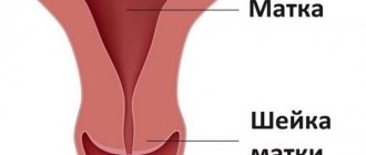
To exclude an ectopic pregnancy, most often the doctor prescribes the first ultrasound examination of the uterus. The most accurate method is a transvaginal examination, which is performed by inserting a sensor into the vagina to determine the location of the fertilized egg (in the uterine cavity, or outside its cavity, which may indicate an ectopic pregnancy).
Sometimes a transabdominal examination is performed, which is performed through the anterior wall of the lower abdominal cavity to determine the thickening or compaction in the walls of the uterus, which is typical for pregnancy.
The first ultrasound examination determines the number of fetuses, SVD (average internal diameter) of the ovum, CTR (coccygeal-parietal size) of the embryo and the exact gestational age. At a period of 4–5 weeks, the size of the fertilized egg reaches from 18 mm to 40 mm (average internal diameter).
fertilized egg
If a doctor, during an ultrasound examination, reports that he sees a fertilized egg in the uterine cavity, the woman can be congratulated, because in 9 months she will become a mother. The presence of a fertilized egg can be determined already on the 7-9th day of missed menstruation.
If the fertilized egg is in the uterus, then the pregnancy is normal, uterine. The specialist will immediately determine the size of the fertilized egg, its shape and location. In addition, he will pay special attention to whether there is a detachment or other pathological conditions.
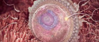
What does a fertilized egg look like?
The fertilized egg is an oval or round body with a diameter of several millimeters. The diameter of the ovum is measured during the first ultrasound. Taking into account its size, a specialist can determine the gestational age. But in some cases the error in determination is 1-1.5 weeks. Therefore, when trying to establish a period, the doctor also takes into account indicators of the coccygeal-parietal size.
At 3-8 weeks of pregnancy
the fertilized egg looks like a formation in the form of a ball or oval. Already at 5-6 weeks, the yolk sac, which provides nutrition to the embryo and performs a hematopoietic function in the early stages of embryonic development, is similar to a bubble inside the cavity of the fertilized egg.
The size of the fertilized egg at this stage of pregnancy is from 1.5 to 2.5 centimeters. It is already possible to examine the embryo at this time. It looks like a five-millimeter strip located next to the yolk sac. And although it is not yet possible to determine which structure and part of the embryo, the heartbeat is already being recorded.
At this time, the baby’s heart beats at a frequency of 150-230 beats per minute.
In addition, the neural tube is already forming in the fetus, and the cells distribute “responsibilities” among themselves, who will create which organs.
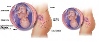
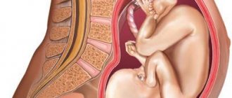
By the end of the 7th week, the embryo has already acquired its characteristic shape in the form of the letter C. At this time, it has already detached from the surface of the fertilized egg. An ultrasound can already distinguish the head, torso and tiny rudiments of arms and legs. An already formed umbilical cord is visible in the fertilized egg.
Irregular shape of the fertilized egg
The normal shape of the fertilized egg is oval or round. If it is flattened on the sides and looks like a bean, this may indicate the tone of the uterus. This condition should be monitored by a doctor.
If nothing bothers the woman, then the deformation does not pose a threat to the pregnancy.
In case of increased uterine tone, doctors prescribe a set of measures (bed rest, medication) to relieve hypertonicity and return the fertilized egg to its correct shape. This can be achieved by relaxing the muscles of the female reproductive organ.
But, if the fertilized egg has an irregular shape, and the woman experiences pain, discharge or symptoms of cervical dilatation, urgent measures must be taken. In such cases, the woman is assigned to the inpatient department of the hospital for safekeeping.
An incipient abortion is called abruption of the ovum. In this case, premature rejection of the fertilized egg from the uterine wall is observed.
Important note: when a spontaneous abortion begins, timely assistance is very important, because, in most cases, the pregnancy can be saved. The main thing is to do everything quickly and competently.
Detachment is accompanied by nagging pain in the lower abdomen, pain in the lower back, and dark red and sometimes brown discharge.
The reasons that cause detachment of the ovum include ovarian dysfunction, various diseases of the woman (tumors, inflammatory processes, infectious diseases), underdevelopment of the genital organs of the expectant mother, severe toxicosis, excessive physical activity, and stress. But the most obvious cause of detachment of the ovum is a lack of progesterone, which is often called the pregnancy hormone.
If a pregnant woman shows signs of abruption of the ovum, she (or relatives) should urgently call an ambulance and call the obstetrician-gynecologist to inform him about what happened. Until the ambulance team arrives, the woman should lie down and raise her legs up. You can rest them against the wall or put them on the back of the sofa.
Detachment of the ovum is dangerous because it can lead to abortion or missed abortion. Therefore, at the slightest suspicion of detachment, you need to seek medical help.
At a very early stage, the embryo in the fertilized egg is not yet visible, and this is the norm. But from five weeks the embryo should already be visualized. If the embryo is not visible, a repeat examination is scheduled after 1-2 weeks. If this time there is neither an embryo nor a heartbeat, they speak of anembryony. In this case, the woman needs to cleanse.
You need to know that even if the ovum is empty, the pregnancy test will still be positive. This is due to the fact that certain mechanisms have been launched in the body, in particular, a special “pregnant hormone” - human chorionic gonadotropin - has begun to be produced.
The reason for the absence of an embryo in the fertilized egg, in most cases, is a failure at the genetic level. Also, anembryonia can be triggered by taking certain medications that are strictly prohibited during pregnancy.
If a woman is diagnosed with an “empty ovum,” which was confirmed by a repeat ultrasound examination, then there is no chance of pregnancy this time.
Then the woman is given the necessary manipulations, prescribed treatment and sent for rehabilitation.
Many women need not only physical, but also psychological rehabilitation to cope with the feelings and emotions that arise as a result of loss.
It is recommended to plan your next pregnancy at least six months later.
Especially for beremennost.net Olga Rizak
Source: https://beremennost.net/plodnoe-yaitso
Weight control
You should start controlling your body weight. But don't go on a diet. Any diet has a very detrimental effect on the baby’s health, since the fetus will not receive the necessary nutrients in sufficient quantities. Remember, fasting and dietary restrictions also affect the mother, and one of the first symptoms will be malaise.
During approximately the first trimester, a pregnant woman gains 1.5 - 2 kg (about 500 grams weekly) in weight.
Every female body is individual, so you should know how much weight you should gain in the fourth week. The calculation is quite simple:
Divide your weight by your height squared. If the resulting number is less than 19.6, you should gain 900 grams, when the number is within 19.6 - 26.0, then 700 grams, and if more than 26, then 500 grams.
As you can see, weight gain varies from person to person. A thin woman should gain more weight than a plump woman in order to give birth to a strong, healthy baby. Therefore, some dietary rules are changing. For example, thin mothers should include more flour and protein foods in their diet to gain weight.
Keep in mind that weight gain numbers can vary depending on the individual, this is not a rule, but only guidelines for the overall picture of how to gain weight correctly. If you have not gained weight within 4 weeks, contact an antenatal clinic or consult a gynecologist.
Deformed fertilized egg in early pregnancy: what does it mean?
Pregnancy is a joyful period for a woman, but it can be overshadowed by various pathologies, for example, improper development of the fertilized egg. With an early ultrasound, the doctor can diagnose changes in its shape or size. What reasons lead to deformation of the ovum, is the pathology dangerous for the mother and baby, what ways can you get rid of the problem?
Causes of deformation of the ovum
At 10-14 weeks of pregnancy, a woman undergoes an ultrasound examination, which allows the doctor to take measurements of the ovum to determine the exact gestational age. Sometimes during the examination a deformed fertilized egg is discovered. The main cause of this pathology is considered to be an increase in uterine tone.
Why does the uterus become toned? Doctors identify several reasons:
- Psychological. The nervous situation in the family and at work, personal fears associated with the course of pregnancy cause stress in a woman. There is general tension in the body, which also affects the condition of the uterus.
- Infectious. Diseases of an infectious nature provoke an inflammatory process that negatively affects the muscle tissue of the uterus and leads to an increase in its tone.
- Intimate life. Violent sexual intercourse during pregnancy actively affects the reproductive organ, causing it to overstrain.
- Hormonal imbalance. Improper hormone production can lead to endometrial growth. Filling the uterine cavity, it negatively affects the surrounding structures, which is how an irregularly shaped fertilized egg is formed.
- Physical activity. Passion for physical exercise provokes tension in the muscle tissue of the uterus and tones it.
During pregnancy, the uterus has heightened sensitivity to any irritants. Even a simple palpation of the abdomen or a short jog can tone it down.
However, these are temporary phenomena and are not considered serious disorders. A cause for concern is the frequent manifestation of increased uterine tone and an elongated outline of the egg.
With such signs, the doctor is forced to constantly monitor the condition of the organ and the course of pregnancy.
Consequences of pathology
During a normal pregnancy, the amniotic egg is located in the uterus. In the short term, at 5-6 weeks, it becomes round or teardrop-shaped. By 6-7 weeks, on a longitudinal ultrasound scan the egg looks oval, but on a transverse scan it remains round. If the examination shows flattening of the ovum on the sides and the absence of an oval shape, then the uterus is in good shape.
What can cause changes in the shape and size of the amniotic egg? A strong increase or decrease in its size indicates the fading of pregnancy, but, as a rule, the doctor continues observations, monitoring the dynamics of the development of changes. An elongated fertilized egg also indicates a frozen pregnancy.
As the tone of the uterus increases, the corners of the egg shell look uneven. In most cases, doctors consider such changes to be harmless unless they are accompanied by dilatation of the cervix, dark discharge and pain. With such symptoms, there is a real threat of fetal loss.
The low position of the fertilized egg in the uterine cavity requires constant monitoring. If it is very close to the cervix, the doctor may decide to remove it. In an ectopic pregnancy, an ultrasound scan shows that the fertilized egg does not contain an embryo; instead, fluid and blood clots are present in the egg cavity.
A sudden acquisition of an irregularly shaped fertilized egg can result in its detachment. Characteristic signs of the pathology are nagging pain and dark red discharge.
The process can be stopped only at the earliest stage of its detection, otherwise everything ends in spontaneous abortion.
Among all the anomalies in the development of the fertilized egg, its detachment from the uterine wall is one of the most serious pathologies.
What to do?
Gynecologists believe that increasing the tone of the uterus does not always have serious consequences. Danger to pregnancy occurs when other symptoms are added to it:
- the cervix dilates;
- pain;
- the appearance of discharge (a characteristic symptom of chorionic detachment).
The patient is prescribed antispasmodic drugs, sedatives and hormonal therapy. During treatment, the deformed structure returns to normal.
If there is a threat of complications, the woman is sent to a hospital to continue the pregnancy (for more details, see the article: How to save a child in the early stages of pregnancy if there is a threat of miscarriage?).
Bed rest and complete peace of mind help to survive an unpleasant period and successfully carry a child.
An important point in prescribing treatment is listening to the fetal heart: if it is present, the chances of success increase significantly. Hospitalization is mandatory if there is a clear threat of miscarriage.
Using various medications, doctors relieve the tone of the uterus, after which the fertilized egg returns to its normal shape. If the diagnosis of a frozen pregnancy is confirmed, curettage of the uterus is performed.
Early fetal death is not a death sentence. In 90% of cases, a woman retains the ability to become pregnant and bear a child. In recurring situations, a deep examination of the body is required to determine the main cause of the problem. If the deformation of the ovum leads to the loss of the fetus, pregnancy planning should be postponed for six months.
The capabilities of modern gynecology make it possible to identify negative changes in the early stages of pregnancy. Minor deformities do not require urgent medical intervention. The prognosis is quite favorable.
Women with this diagnosis usually feel normal during pregnancy and give birth successfully.
If the ultrasound shows a problem, try to strictly adhere to the doctor’s recommendations and follow them strictly.
Source: https://www.OldLekar.ru/beremennost/plod/deformirovannoe-plodnoe-yajco.html
Symptoms
The 4th week definitely gives signs of pregnancy, because the fertilized egg has implanted in the uterus and the development of the embryo has begun. But still, more often than not, a woman does not notice severe symptoms or confuses them with signs of the onset of menstruation.
What sensations arise?
- The lower abdomen hurts and pulls, due to the body’s restructuring to perform new functions.
- The mammary glands swell and prepare to feed the baby, which leads to pain and breast enlargement.
- In individual cases, early toxicosis begins.
Toxicosis is a reaction of the brain to the process of changes in metabolism in the body, in connection with the birth of a new life. Signs of toxicosis are similar to those of food poisoning: nausea, vomiting.
Fighting toxicosis
Deformed fertilized egg in early pregnancy
Hello, dear blog readers!
Have you heard that the fertilized egg can have an irregular shape? I also didn’t know that such a pathology existed. Today we will talk to you about what a deformed fertilized egg is in the early stages of pregnancy and why this happens. But the main thing is to figure out whether this condition is dangerous and how to avoid it.
Is shape important?
As soon as a woman realizes that she is pregnant, she is required to register with a doctor so that the specialist can monitor the condition of the mother and her child. In the first trimester, various studies are required, including ultrasound.
The specialist first determines the presence of pregnancy, which will be indicated by the presence of a fetal element - this is the embryo and the various membranes surrounding it. It is diagnosed already at 5 weeks of age. Then the pathology is determined.
In studies, the main indicator is the average and internal diameter of the ovum (SVD). The gestational age is judged by the size of the existing fertilized egg, which in the first weeks should be oval or spherical in shape. This is the norm.
But sometimes the doctor sees it as too large or elongated; this is necessarily recorded in the history of the pregnant mother. Should I worry about this diagnosis? As practice shows, this does not apply to pathological conditions, provided that it is not accompanied by other symptoms.
Therefore, the prognosis for pregnancy in the case of a deformed fetal element is almost always positive. But it will be possible to say more accurately only after conducting certain studies.
Why is this happening
Mommies are most interested in the question of why her fertilized egg may be deformed in the early stages. In fact, there may be several causes of pathology. The main one is deformation due to severe hypertonicity of the uterus. This affects the shape of the fertilized egg, which is compressed.
Other risk factors include:
- suffered stress;
- emerging infections in the reproductive system;
- viral and bacterial infections in other organs;
- hormonal imbalance.
This is not a complete list of reasons. What does this mean for a woman? A full range of studies, after which the doctor usually prescribes medication. But mommy herself will need to behave carefully, otherwise the consequences may be unpredictable.
What to do? Or rather, what you shouldn’t do is make love to your husband, overload your body with physical exercise, or worry about various trifles.
What else will the study show?
Ultrasound shows the doctor not only the shape, but also the pathologies that can be caused by this. For example, already at 6 weeks you can predict the fading of pregnancy.
Pathologies can be different:
- small ovum;
- large egg size;
- elongated shape.
We should talk about a small size if the egg does not correspond to the gestational age. But most often this is not a pathology, but an error in determining the week of pregnancy. But the same can be observed in the presence of a frozen pregnancy.
In this situation, it will be necessary to conduct additional research. But if a woman also has brown discharge, then she should rush to get tested. This may be due to a missed or ectopic pregnancy. Delay is unacceptable!
A large egg also indicates an abnormally developing pregnancy. Research shows that it develops without an embryo. But in this case, mistakes are often made during ultrasound examination, so it is best to carry it out after the 7th week of pregnancy.
Normally, the shape can be round or oval. But sometimes an ultrasound shows an elongated egg. Usually we are talking about uterine hypertonicity, which requires treatment in a hospital. An elongated egg is observed in twins and this does not indicate pathology, this is the norm.
A damaged ovum during twins often results in the loss of one embryo. The second can develop further in the absence of other pathologies.
From embryo to fetus
Women are often interested in why, when they talk about improper development of the egg, they don’t mention the fetus? It's simple. Until the 8th week, obstetricians do not call the new life in a woman’s body a fetus, since it is not exactly a child, but a certain substance that will very soon turn into a baby. But every week it develops, increasing in size.
This allows you to determine the shape of the developing fertilized egg week by week:
- up to 16 weeks - 1 mm per day;
- until the end of pregnancy - up to 2.5 mm per day.
The 6th week is important, at this time the digestive system and spleen are born. With a size of 16 mm - the stomach and esophagus. It is at this time that they say that a woman is carrying a child. And this is a difficult period associated not only with the restructuring of the entire body, but also with the discomfort that the woman experiences during this process.
And each of us wants to look as attractive after a successful birth as before the “interesting situation.” And mothers will be helped with this by a special cream for stretch marks for pregnant women – “Babycoccole”. The volume of 300 ml allows you to use the composition for a long time.
And its components are completely safe for a woman carrying a child. Before use, it is recommended to take a shower and apply a small amount of cream to the area prone to stretch marks. Usually this is the stomach, chest, thighs.
Is the pathology treatable?
As a rule, a deformed fetal element at a very early stage does not affect the woman’s well-being. But if such a condition is detected during the study, the doctor must immediately take action. Tests are required, including tests for progesterone, to rule out hormonal imbalance.
If this diagnosis is confirmed, the gynecologist prescribes Duphaston or Utrozhestan. Reviews about the drugs are the best, which indicates the high effectiveness of the products. As for the dosage, it is always individual and determined by the degree of hormone deficiency.
If the cause is uterine hypertonicity, then antispasmodics are prescribed, the most popular ones include:
In addition, medications with magnesium and iron and sedatives are prescribed to relieve tension. With timely and proper treatment, the pathology can be dealt with by the 13th week of pregnancy or a week or two later. But treatment must be carried out under strict medical supervision.
If necessary, you need to go to the hospital. Here it is important not only to follow the doctor’s recommendations, but also to feel comfortable. In the latter, women are helped by a set - a robe with a “Mamalandia” shirt. Very comfortable clothes for staying at home and in the hospital.
You can wear a shirt, and if you need to go out, just throw a robe on top and tie it with ribbons. No buttons, rough locks! The kit is designed for any stage of pregnancy, thanks to just this mounting option. Clothes will also be indispensable when feeding your baby.
Friends!
As you have learned, a deformed ovum is a completely understandable and, most importantly, not dangerous condition in some cases. But prompt research and treatment are important.
Tell your friends about this on social networks, I’m sure the article will help avoid panic. Subscribe to the blog and you will be among the first to receive news and updates. This way you can avoid common mistakes during pregnancy. Until new topics!
Sincerely, Tatyana Chudutova, mother of three wonderful children!
Source: https://vladimir-sport.ru/vse-o-detyah/beremennost-i-rody/plodnoe-yajco-deformirovano.html
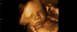
Photo of the fetus on ultrasound
So, the long-awaited 4th week of pregnancy has arrived. Now you know for sure that you are pregnant and enjoy watching the changes in your body.
Empty fertilized egg (anembryonia) during pregnancy: causes, signs
Today, such pathology as anembryonic pregnancy (anembryonic pregnancy) or an empty fertilized egg is becoming more and more common. Anembryony is the absence of an embryo in the fertilized egg. There are membranes, amnion (water membrane), chorion (villous membrane), but no unborn child (embryo).
Some of these pregnancies are interrupted at a very early stage before implantation into the uterine wall - this is how the body gets rid of the defective embryo. But if the fertilized egg manages to attach to the wall of the uterus, then its chorion begins to produce hCG, as in normal pregnancy (see “HCG during pregnancy”).
Therefore, with anembryonia, a woman experiences a delay in menstruation, and there may be signs of pregnancy such as nausea, changes in taste, fatigue, and breast swelling. Since pregnancy tests are based on hCG detection, they will be positive.
However, anembryonic pregnancies cannot develop normally and always end in miscarriage in the first trimester. Often a miscarriage occurs before a woman even finds out that she is pregnant or in the first days after a missed period.
Empty fertilized egg (anembryonia) causes
The main reason for the appearance of anembryonia or an empty fertilized egg is chromosomal and genetic abnormalities. Most often they arise spontaneously at the time of cell division, that is, in the first days after fertilization. During the division of the cells responsible for the formation of the embryo, a malfunction occurs and the embryo is not formed, while other cells continue to develop and create an empty fertilized egg.
There is another situation when the embryo is formed, but dies in the early stages of pregnancy and it is so small that it is not visible on ultrasound.
Signs of anembryonia
Pregnancy with an empty sac may have the same symptoms as a normal pregnancy. However, with anembryonia, spotting and spotting is often observed, as the uterus tries to get rid of the defective fertilized egg.
An empty ovum can only be determined using ultrasound. However, all women are not recommended to undergo an ultrasound in the early stages; such an examination should be carried out only when indicated, for example, in the presence of bloody discharge or severe abdominal pain.
An ultrasound performed too early can give an erroneous result and add unnecessary stress to a pregnant woman's life. There are often cases when the diagnosis of “anembryonia” turns out to be false. An ultrasound was performed very early or the gestational age turned out to be shorter than expected, and the small embryo was simply not seen.
It is possible to determine anembryonia by ultrasound no earlier than 5-6 weeks of pregnancy, but even at this period there may be errors.
Anembryonia: what to do
If anembryonia is suspected, one should not rush to terminate the pregnancy. Firstly, the diagnosis may be false. It is imperative to conduct another ultrasound examination in about 7-10 days.
Secondly, if the diagnosis is confirmed, it is recommended to wait 2-3 weeks. Cleaning is required in very rare cases; most often, an empty fertilized egg comes out of the uterine cavity on its own. Basically, spontaneous termination of pregnancy with an empty ovum occurs up to 8 weeks.
Only in some cases may instrumental removal of the fertilized egg be necessary. It is also possible to medically terminate an anembryonic pregnancy, when a woman takes certain medications and the fertilized egg comes out on its own.
It is worse when, when bleeding occurs, the doctor prescribes progesterone and other drugs to maintain pregnancy and this prevents the evacuation of the defective fertilized egg. In such cases, the likelihood of surgical termination of pregnancy (cleaning) increases.
After a pregnancy loss with an empty sac, your doctor may recommend waiting one to three menstrual cycles before trying to conceive again.
Pregnancy after anembryony
In most cases, anembryony occurs accidentally due to defective conception and does not recur. If the empty ovum repeats more than two times in a row, the couple needs to be examined. A woman needs to start with a thyroid examination. The thyroid gland can produce antibodies that penetrate the embryo and lead to its death.
Repeated anembryonia can occur due to pathological sperm, so a man must have a spermogram. After three pregnancy losses, the couple is also advised to undergo genetic counseling.
It is important to understand that you are not to blame for anything and there is nothing you could have done to prevent this loss. An empty fertilized sac occurs spontaneously due to improper cell division or a poor-quality egg or sperm. This pregnancy has no future, so your body rejects it.
It is impossible to prevent anembryony. Anembryonia can occur at any age in completely healthy women. If you do not interfere with the natural course of events and do not take medications that preserve pregnancy, then anembryonia in most cases ends with spontaneous termination in the early stages and does not affect subsequent pregnancies.
Have you lost your desired baby due to anembryonia? Do you find it difficult to cope with mental pain? We recommend reading the article “How to survive a miscarriage or frozen pregnancy,” and you can also preserve the memory of your child by visiting the “Memory Page.”
Source: https://nashy-detky.com.ua/beremennost/nevynashivanie-beremennosti/pustoe-plodnoe-yajco-anembrioniya-pri-beremennosti-prichiny-priznaki.php
Nutrition
It's time to switch to healthy eating. Avoid fatty foods, eat more vegetables and fruits, but do not indulge in exotic species (they cause diabetes in a child).
Doctors recommend drinking folic acid in the first trimester to help the body preserve the fetus and improve the development of the baby at the beginning of pregnancy.
Try to remove coffee from your diet. It suppresses appetite and causes uterine tone. Coffee excites, which affects a woman’s normal sleep; her body gets stressed, which negatively affects the fetus.
Consume dairy products, fish and lean meat. Avoid eating foods that are low in nutrients (cakes, pastries) but lead to weight gain.
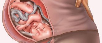
Initial stage of pregnancy: fertilized egg
Externally, the fertilized egg resembles a round or oval egg, the size depends on the stage of pregnancy. It is the outer shell in which the fruit is located and develops, hence the name. After fertilization, the egg moves to the uterus. During the promotion period, an outer shell is created, which not only protects the fetus, but also provides nutrition at the initial stage.
Why this name
A fertilized egg is called an egg because its shape and structure repeats the structure of a standard egg:
- shell;
- amniotic fluid;
- yolk sac;
- embryo
Over time, the egg transforms inside the uterus. The outer shell, called the chorion, has villi that serve to anchor it to the mucous membrane of the uterus. Later, the placenta is obtained from the chorion.
Amniotic fluid (analogue of egg white) is a nutrient medium produced by the amnion, that is, the inner membrane of the fertilized egg.
The yolk sac is the nutritional reserve that, when pregnancy occurs, the egg feeds the future embryo. It connects to the embryo in the area of the umbilical cord, and through a small tubule transfers nutrients to the zygote.
For now, you can only see on an ultrasound what the fertilized egg looks like.
How the fertilized egg is formed
After two cells, male and female, have merged, they continue to divide. In this case, the entire structure moves towards the uterus.
By the time it enters the uterine cavity, this formation will consist of 32 cells, covered with a membrane on top, which is called chorion. The entire structure is called a fertilized egg or blastocyst.
Its shell is covered with specific villi, with which it will later attach to the wall of the uterus for further development.