Pathogenesis . The normal vitelline duct is obliterated at the 7th week of embryonic life. If it does not heal after the umbilical cord falls off, a fistula is formed in the navel area, the discharge from which depends on the nature of the pathology.
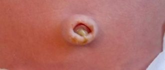
Symptoms, course. With complete non-closure of the vitelline duct, a small bright red, corolla-shaped s. is usually visible at the site of the fallen umbilical cord. a hole in the center, a granuloma-like formation from which mucus and sometimes gas bubbles or feces are constantly released. The probe can be inserted through the hole quite easily. A patent urinary duct is less common. In this case, urine is constantly or periodically (when the baby cries) released from the navel.
Recognition . An umbilical fistula must be differentiated from a granuloma of the pelvis. In cases where the umbilical wound does not heal for a long time and there is a constant discharge from it, you should think about an umbilical fistula.
Treatment is surgical.
Congenital hernia of the umbilical cord is a developmental defect
Pathogenesis . In a certain period of embryonic development (up to 6-10 weeks), part of the intestine is located outside the abdominal cavity, and after birth remains in the cavity of the umbilical cord.
Symptoms, course . Through a significant defect in the abdominal wall in the umbilical region, the abdominal organs protrude in the form of a tumor. The hernial sac is covered with a thin transparent membrane of the amnion. The skin is missing. The liver, stomach, intestines, and spleen can exit into the hernial sac.
Treatment . Urgent surgery. For small hernias, the contents are easily reduced; treatment is carried out conservatively, using bandages.
Calcification of the umbilicus
Navel calcification is the deposition of lime in the area of the umbilical wound or along the umbilical vessels. In recent years, I began to meet quite often. The history sometimes indicates the introduction of calcium chloride into the umbilical vessels.
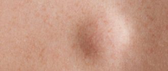
Symptoms and course. In the navel area, compaction is determined due to calcium deposition. The vessels are poorly obliterated, and therefore bleeding from the arteries may occur.
Treatment . Removing calcification with tweezers or a sharp spoon. If this fails, it is removed surgically.
What kind of pathology is navel fungus in newborns, why does it occur, how to treat it
Navel fungus is not a specific disease and not an infectious disease .
Rather, it is an individual feature of the newborn’s body, the reasons for the development of which are not fully understood. A wide umbilical ring often becomes the source of granuloma formation. When the umbilical cord falls off, the resulting cavity is filled with fungus. The appearance of the granuloma is a mushroom-shaped growth, at the base of which there is a stalk. The fungus occupies the entire area of the umbilical fossa or can extend beyond its boundaries.
Granuloma not only causes aesthetic discomfort, but also often causes negative consequences. Complications develop against the background of secondary infections entering the umbilical ring.
If infection is not attached, fungus proceeds without disturbing the general condition or unpleasant clinical manifestations. Changes in blood parameters are not diagnosed. Complete treatment eliminates granuloma in a short time.
Reasons for appearance
As noted above, pathology forms after the umbilical ring falls off. A characteristic growth forms in its cavity, which does not cause discomfort to the child.
The reasons for this are as follows:
- the presence of a wide, thick umbilical cord or a large umbilical ring (after the remainder falls off, granulation tissue forms on the free one);
- entry into the umbilical wound of a secondary infection - the source of granuloma formation or its complications;
- birth of a child prematurely (premature children are diagnosed with pathology many times more often than children born on time);
- if the baby is heavy at the time of birth.
This is not an exhaustive list of reasons why granuloma develops. Each body is individual and reacts differently to certain provoking factors. In addition, fungus is often confused with other, more dangerous diseases.
Symptoms
This is what navel fungus looks like
Umbilical granuloma in a newborn is not characterized by obvious symptoms.
The child behaves as usual.
Local manifestations include the presence of characteristic growth and redness in this area.
The neoplasm has a dense consistency and a bright pink color.
If an infection occurs, this process occurs with an increase in general temperature and loss of interest in food. As a result, weight decreases and weight gain stops. The child becomes whiny, restless, and general malaise may occur.
Local symptoms include the following:
- discharge from the umbilical ring of a characteristic exudate that is mucous, purulent or bloody in nature;
- redness of the affected area;
- increased local temperature;
- swelling of the skin in the area of the umbilical ring;
- if an inflammatory process develops in the lymph vessels, there are red stripes.
Umbilical fungus in newborns is not difficult to diagnose. The situation becomes more complicated when omphalitis occurs, because this pathology occurs in several forms, each of which has its own clinical symptoms :
- Catarrhal . Diagnosed in most cases. A light mucous exudate is released from the navel. Sometimes there are blood streaks in it. The skin is slightly reddened. There are no complications. Catarrhal omphalitis is easy to treat.
- Purulent . A light yellow or brown exudate of a purulent nature, viscous consistency and with an unpleasant odor is released from the umbilical ring. The skin is swollen and red. The general temperature has increased to low-grade levels. General well-being is disrupted and anxiety increases. A favorable prognosis is observed with complete and timely treatment.
- Phlegmous . It is characterized by the severe general condition of the newborn, an increase in general temperature to 39-40 degrees. Interest in food is completely lost. The umbilical ring is filled with an ulcer that contains purulent masses. The infection involves the surrounding tissue. Local and systemic drugs are used as treatment.
- Necrotic . Infected tissues die, and an inflammatory process develops in the peri-umbilical vessels. The baby's general health is poor. This form of the disease occurs as a result of delayed consultation with a doctor.
Stages of healing of the umbilical wound
Umbilical granuloma has pronounced symptoms and therefore can be diagnosed during the initial examination .
If such a pathology is suspected, the mother and child should visit a pediatrician or pediatric surgeon.
Additional diagnostic measures are prescribed if there is a suspicion of the development of infectious omphalitis.
To determine the type of pathogen, biomaterial isolated from the umbilical wound is taken, which is sent for laboratory diagnostics. Using the results of tank culture, the level of sensitivity of the pathogenic microorganism to the drugs used (usually antibiotics) is determined.
Ultrasound examination can identify complications . This may be an abscess or phlegmon of the abdominal region. If necessary, an X-ray examination is prescribed, which also helps to identify the negative consequences of the disease. The general condition of the body can be determined by performing a general blood and urine test.
How to treat the disease
Treatment of navel fungus in newborns is not carried out as such . The baby’s general condition returns to normal on its own without taking additional measures, and the fungus heals after a short time.
Despite the ability to self-stop, granuloma sometimes requires specific therapy. Therefore, you should not miss the first warning symptoms, but you should consult a doctor if:
- the occurrence of edema;
- redness around the umbilical ring;
- presence of exudate.
Every mother can treat fungus independently at home. In combination with the use of medications, it is necessary to maintain daily hygiene of the affected area. It is recommended to put on and remove the diaper carefully, avoiding injury to the granulosa tissue. Which diaper to choose for a newborn is described in the article at the link.
For a speedy recovery, the following drugs are used:
| Name, release form | Dosage, method of application | Contraindications | Side effects | Average price in rub. |
| HYDROGEN PEROXIDE solution | Apply 2-3 drops after bath | Hypersensitivity | Allergic reaction | 3-10 |
| BRILLIANT GREEN solution | Treat granuloma after a bath | Hypersensitivity | Allergic reaction | 5-14 |
| IODINE solution | Treat after bath | Hypersensitivity | Allergic reaction | 10-15 |
| CHLOROPHYLLIPTE, solution | Treat the navel several times a day | Hypersensitivity | Allergic reaction | 60-100 |
- Hydrogen peroxide is an environmentally friendly substance, which makes it available for use by both adults and children, including newborns. The drug has a disinfectant effect, preventing infection from entering the wound.
- DIAMOND GREEN solution is used for many pathologies of the skin, including open wounds of the epidermis, abrasions and cuts. For fungus, the medicine cannot be used together with other disinfectant preparations, which contain iodine, alkalis and chlorine.
- IODINE has a disinfecting effect. Helps reduce the intensity of the inflammatory process and eventually stop it completely. In addition to allergies to substances, prohibited use includes trophic and diabetic ulcers on the skin.
- CHLOROPHILLIPT is used even when local antibiotic drugs are ineffective, to which staphylococci may be resistant. Approved for use by both adults and newborns.
Prevention
Do not forget to treat the umbilical wound with hydrogen peroxide and brilliant green until it is completely healed
It is almost impossible to prevent the formation of granulomas , because this is an individual feature of the body.
You can prevent the addition of a secondary infection, and this is:
- maintaining personal hygiene and daily treatment of the navel with appropriate medications (read more about this here);
- eliminate friction of clothes and diapers on the navel;
- thoroughly wash toys, iron clean things, wash your baby’s hands or treat them with a disinfectant;
- Clean the living area daily.
Complications and prognosis of the disease
The only complication of navel granuloma in newborns is the development of the infectious disease omphalitis, which occurs against the background of an infection. If this complication is left untreated, the risk of blood poisoning increases. When granulation tissue grows rapidly, surgery may be required.
conclusions
If there are no complications, fungus quickly passes without the possibility of relapse. The prognosis is favorable. Dr. Komarovsky tells how and what to treat the umbilical wound.
Magazine subscription
Source: https://pupsek.com/zdorov-e-rebenka/detskie-bolezni/fungus-pupka-u-novorozhdennyh.html
Omphalitis
The disease is most often caused by streptococcus, staphylococcus, and less commonly by intestinal, pseudomonas or diphtheria bacillus. Along with antibiotic therapy, it is important to increase the resistance of the newborn’s body with good care, proper breastfeeding, administration of γ-globulin, hemotherapy, blood transfusions, and vitamin therapy.
Prevention . Asepsis when ligating the umbilical cord and caring for its remnant and umbilical wound. Accelerated falling off of the umbilical remnant, clamping of the umbilical cord with a Kocher clamp, and treatment of the umbilical cord remnant with an alcohol solution of gramicidin (1: 100) promote faster epithelization of the umbilical wound and prevent its infection.
Omphalitis in adults and children: treatment of navel inflammation
This is a multifactorial disease that affects the navel area, skin and subcutaneous layer. This can severely damage the lymphatic and blood vessels in the body.
Most often, navel omphalitis affects a newborn in the first days of life. As a rule, the disease occurs with complications. This is due to the transition of processing the umbilical cord stump and preventing infection.
Omphalitis in adults; treatment of its symptoms requires timely consultation with a doctor.
Definition of disease
A rather rare occurrence of navel disease in adults. This disease occurs in a mild form, and adequate and modern treatment brings results in a short period of time.
Omphalitis of bacterial origin is a surgical pathology; with the development of more severe pathology, omphalitis sometimes requires surgical intervention.
To avoid surgery, you must pay attention to the main symptoms and treat inflammation.
Omphalitis is a fairly simple disease, but its main types and forms are quite serious. These are inflammatory processes in the skin that result in the development of a wound.
Based on their origin, two types of lesions :
- Primary - the development of infection directly in the umbilical wound itself;
- Secondary - the addition of infection to the fistula (due to the progression of the disease).
Nature of the inflammatory process:
- Catarrhal (serous-purulent, simple and “weeping navel”) is the more common form. Occurs when epithelium begins to slowly cover the wound. During this process, a colorless liquid comes out of the wound, and then granulation and blood crusts appear.
- Phlegmonous.
- Necrotic (gangrenous) - this type is very difficult to cure.
- Purulent lesions are a particularly serious form of omphalitis, which causes severe ulcers, purulent discharge, and also protrudes the navel above the abdominal cavity.
According to forms, it is divided into acute, chronic and bacterial nature of origin.
Causes of navel infection
A factor in the development of navel damage is considered to be an infection that affects the body (staphylococcal or E. coli), which penetrates the umbilical wound. It heals completely after the baby is born. The pathology affects newborns, as well as older children and even adults. General factors that provoke the development of infection:
- failure to follow basic hygiene rules, which very often leads to the spread of infection, causing not only children, but also adults to become infected;
- poor wound treatment;
- contamination by urine or feces;
- dirty bed linen, underwear or towel.
It is also important to consider pregnancy, during which many infections are transmitted from mother to newborn baby. If she has an infectious disease, bacteria can enter the umbilical cord.
Symptoms of damage to a newborn
For infection to develop, evidence of inflammation . In newborns, these factors include:
- decreased immunity, a vulnerable and thin layer of epithelium, due to prematurity, low weight or immaturity of the baby’s body;
- too loose subcutaneous fat layer with omphalitis in children with high weight;
- catheterization of the umbilical cord vessels, if it was carried out without observing sanitary standards (during resuscitation, infusion therapy, the need for blood transfusion and other medical procedures);
- infectious skin lesions (phlegmon, pemphigus, vesiculopestulosis, and erysipelas);
- purulent lesions of internal organs (pneumonia, meningoencephalitis and osteomyelitis);
- non-compliance with the thermal chain (the baby is not laid on the woman’s stomach), as well as the lack of contact between mother and baby after the birth process;
- a large number of doctors and nurses nearby, medical staff not following sanitary standards;
- treatment of the remaining umbilical cord using alcohol antiseptics without the testimony of the attending physician;
- poor child support and living conditions in the house, unfavorable social situation;
- bacterial infection during pregnancy, as well as during childbirth;
- long-term falling off of the umbilical cord stump with congenital hypothyroidism;
- the presence of complete and incomplete fistula.
Source: https://kozha.me/zabolevaniya-kozhi/omfalit
Periarteritis of the umbilical artery
Pathogenesis. Develops when asepsis is not observed when ligating the umbilical cord. The umbilical wound has no visible changes. On palpation below the navel, the umbilical arteries are identified as a compacted cord. The disease can be latent.
Treatment . Compresses with a hypertonic solution of sodium chloride (10%) or rivanol (1: 1000), ensuring the outflow of pus from the artery. Broad-spectrum antibiotics. Hemotherapy, γ-globulin. Plasma transfusion. For peritonitis - surgical intervention.
Cutaneous belly button - excess skin of the abdomen extends onto the umbilical cord. Refers to a cosmetic defect. No treatment required.
Umbilical bleeding occurs when the ligature is poorly applied to the umbilical cord, as well as when normal obliteration of blood vessels is delayed, or when there are bleeding granulations. Treatment. Pressure bandage, vitamin K, in case of significant bleeding, ligation of blood vessels.
Phlebitis of the umbilical vein develops when the umbilical vessels become infected if antiseptic rules are not followed when tying the umbilical cord or performing a replacement blood transfusion in a newborn with hemolytic disease. Symptoms, course. The umbilical wound may remain unchanged. Complication: sepsis. Treatment. Broad-spectrum antibiotics, blood transfusions, γ-globulin, B vitamins (B6, B12). Feeding with mother's milk. Proper care.
INFECTIOUS DISEASES OF THE UMBILICAL WOUND, UMBRICAL CORD AND UMBILIAL VESSELS
Omphalitis is an inflammatory process of the bottom of the umbilical wound, umbilical vessels, skin and subcutaneous tissue in the navel area.
Classification. There is no accepted classification. Based on clinical and morphological data, the following forms of omphalitis are distinguished: catarrhal omphalitis (weeping navel), navel fungus, purulent, phlegmonous and necrotic omphalitis. When the umbilical vessels are affected, they speak of phlebitis and arteritis.
Etiology. Among the pathogens that cause inflammation of the umbilical wound are both gram-positive microorganisms (staphylococci, streptococci) and gram-negative ones (Escherichia coli, Proteus, Pseudomonas aeruginosa, etc.). The cause of gangrene of the umbilical cord is anaerobes.
Pathogenesis. The pathogen penetrates the tissue adjacent to the navel transplacentally, through the umbilical cord stump, causing productive, purulent or necrotic inflammation. The infection spreads and is fixed in the umbilical vessels. The incidence of phlebitis in newborns is increased by catheterization of the umbilical vein. The spread of inflammation leads to the development of phlegmon in the navel area. With thrombophlebitis of the umbilical vein, the infectious process along the portal vein can spread into its intrahepatic branches with the formation of purulent foci along the veins even after the umbilical wound has healed.
Clinic. The most common and prognostically favorable form of the disease is catarrhal omphalitis (weeping navel)
, in which a long-term non-healing granulating wound appears at the bottom of the umbilical wound with scanty serous discharge, periodically covered with a crust.
Granulations can grow excessively, forming a mushroom-shaped protrusion ( umbilical fungus
). The umbilical vessels are not palpable. The general condition of the child is satisfactory, body temperature is normal, there are no changes in the peripheral blood.
If there is purulent discharge from the umbilical wound, swelling and hyperemia of the umbilical ring, they speak of purulent omphalitis
. In some cases, inflamed umbilical vessels (elastic cords above or below the navel) begin to be palpated. The disease may be accompanied by intoxication, increased body temperature, and inflammatory changes in the blood.
Phlegmonous omphalitis
occurs as a result of the spread of the inflammatory process to the umbilical area. With this form, swelling, tissue infiltration, skin hyperemia, and protrusion of the umbilical region appear. In some cases (if the crust is not removed when treating the umbilical wound), an ulcer with undermined edges and fibrinous deposits forms at the bottom of the umbilical wound. Deterioration of the condition, lethargy, weak breastfeeding, regurgitation, pallor of the skin or its pale gray tint, an increase in body temperature to febrile levels, a decrease or absence of weight gain are noted.
Necrotizing omphalitis -
an extremely rare complication of the phlegmonous form of omphalitis in premature and severely weakened children. The process extends deeper. The skin acquires a purplish-bluish color, necrosis and detachment from the underlying tissues occur. This creates a large wound. The muscles and fascia in the abdominal wall are quickly exposed. Intestinal eventration may subsequently occur. External manifestations of the inflammatory process resemble necrotic phlegmon of a newborn. This form of omphalitis is the most severe and often leads to the development of sepsis.
With thrombophlebitis of the umbilical vein
an elastic band above the navel is palpated.
For thromboarteritis
The umbilical arteries are palpated radially below the umbilical ring.
In case of development of periphlebitis and periarteritis
the skin over the affected vessels is swollen and hyperemic, there may be tension in the muscles of the anterior abdominal wall, which can be determined by palpation (positive Krasnobaev’s sign). With light massaging movements from the periphery of the affected vessel to the umbilical ring, purulent discharge appears at the bottom of the umbilical wound. In a number of cases, the symptom of a “secondary opened navel” develops, when hemorrhagic discharge reappears from the umbilical wound, after its epithelization has already taken place. Possible development of intoxication.
Gangrene of the umbilical cord (umbilical cord)
develops in the first days of life. The mummification of the umbilical cord remains stops, it becomes moist, acquires a dirty brown tint and an unpleasant putrefactive odor. As a rule, the development of sepsis is noted.
Diagnosis of omphalitis –
clinical and is established in the presence of purulent or serous inflammation of the umbilical wound and umbilical vessels with purulent or serous discharge, infiltration and hyperemia of the umbilical ring, palpable umbilical vessels, delayed epithelization of the wound.
Laboratory research.
In a hemogram in severe forms of omphalitis, leukocytosis with neutrophilia and a shift of the leukocyte formula to the left to young forms can be detected, and an increase in ESR is possible. Bacteriological examination of blood and discharge from the umbilical wound allows one to clarify the etiology, and an antibiogram allows one to prescribe adequate etiotropic therapy.
Instrumental research.
In some cases, when the umbilical wound is wet for a long time, probing is performed to exclude incomplete umbilical fistulas. To exclude urachus, a test is carried out with the introduction of an aqueous solution of methylene blue into the bladder or fistula.
Differential diagnosis. A weeping navel must be differentiated from umbilical fistulas (incomplete umbilical fistula, urachus and complete intestinal fistula), leading to prolonged weeping of the umbilical wound. Phlegmonous and necrotic omphalitis are differentiated from phlegmon of newborns and erysipelas.
About phlegmon
we can say when the inflammatory process extends far beyond the umbilical ring. The skin has a purplish-cyanotic tint, its blood supply is disrupted, cyanotic areas alternate with pale ones, and a fluctuation occurs in the center. Subsequently, signs of necrosis appear with the formation of a demarcation line.
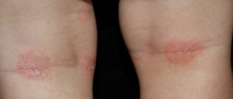
Erysipelas
is an independent disease and is not associated with omphalitis, although the umbilical region is a typical location for this disease. Newborns often have an erythematous form of erysipelas. A bright purple spot without clear contours appears on the affected skin. Hyperemia spreads unevenly, in the form of “tongues of flame.” The skin is shiny, tense, displaced in relation to the subcutaneous tissue, warm to the touch. Later, the color of the skin becomes normal, leaving a slightly peeling surface. Blisters, subcutaneous abscesses, and necrosis may occur.
Navel fistulas
can be complete or incomplete. A complete fistula is caused by non-closure of the duct between the navel and the loop of intestine, or by the preservation of the urinary duct. The main manifestation of a complete fistula is prolonged weeping of the umbilical wound (sometimes the release of intestinal contents).
If the contents of the umbilical wound are sour, a patent urinary duct may be suspected.
For incomplete navel fistulas
(non-closure of the distal urinary or bile ducts) the clinical picture of catarrhal omphalitis develops.
If umbilical fistula is suspected, consultation with a pediatric surgeon is indicated. The final diagnosis is established after fistulography or a methylene blue test).
Treatment. Objectives of treatment: sanitation of the umbilical wound, detoxification, immunocorrection.
Indications for hospitalization.
For catarrhal omphalitis and navel fungus, with active patronage and good social conditions in the family, hospitalization is not necessary. For purulent omphalitis, hospitalization is indicated in the presence of intoxication, involvement of the umbilical vessels in the process, as well as children at risk for generalization of the infection and unfavorable microsocial conditions. For other forms of omphalitis and inflammation of the umbilical vessels, the child must be hospitalized.
Non-drug treatment.
Hygienic baths with a solution of potassium permanganate 1:10000, decoctions of a series of herbs, chamomile flowers, and celandine herbs are shown. In severe cases, skin cleansing is carried out using wet wipes. Physiotherapeutic methods (ultraviolet irradiation) are widely used in treatment.
Drug therapy.
Local therapy: depends on the form of the disease, the nature and extent of the local process. For catarrhal and purulent omphalitis, the umbilical wound is treated with a 3% solution of hydrogen peroxide, then a 5% solution of potassium permanganate, or a 2% alcohol solution of brilliant green. You can use powder with bacitracin and neomycin (baneocin), treat the umbilical wound with antiseptic solutions (chlorophyllipt, 10-15% propolis solution, 1% solution of eucalyptus leaf extract, etc.). Ultraviolet irradiation of the umbilical wound is used. In case of navel fungus, the umbilical wound is treated by cauterizing granulations with a 5% solution of silver nitrate by a doctor. For the phlegmonous form of omphalitis, bandages with a solution of dimethyl sulfoxide, with ointments on a hydrophilic basis (levosin, levomekol), with hypertonic solutions of 5-10% sodium chloride solution, 25% magnesium sulfate are used. In case of necrotizing omphalitis and gangrene of the umbilical cord after surgery, the wound is treated openly using hydrophilic ointments (see above). For phlebitis and arteritis of the umbilical vessels, the umbilical wound is cleaned, similar to a weeping navel and purulent omphalitis, as well as dressings with 2% troxerutin gel.
When using film-forming drugs (Lifuzol, etc.) approved for treating the umbilical wound in an obstetric hospital, in cases of signs of omphalitis, the film is removed with 70% ethyl alcohol; Subsequently, treatment of the umbilical wound is carried out as indicated above.
General treatment is described in the section Pemphigus of newborns.
Surgery.
Surgical treatment is indicated in case of abscess formation with phlegmonous omphalitis. In case of necrotizing omphalitis and gangrene of the umbilical cord, necrectomy must be performed.
Forecast. Favorable for mild forms of omphalitis, inflammation of the umbilical vessels, subject to timely and adequate therapy. Phlegmonous and necrotic omphalitis, gangrene of the umbilical cord with complications (including sepsis) can be fatal.
| Questions for the exam. Infectious and inflammatory diseases of the skin and subcutaneous tissue. Vesiculopustulosis. pemphigus, abscesses, exfoliative dermatitis, candidiasis of the skin and mucous membranes, panaritium, paronychia, phlegmon. Etiology. Clinical picture. Diagnostics. Differential diagnosis. Treatment. Rational choice of antibiotics. Diseases of the umbilical cord, umbilical wound and blood vessels. Omphalitis, thrombophlebitis, arteritis of the umbilical vessels, gangrene of the umbilical cord. Etiology. Clinical picture. Diagnostics. Differential diagnosis (fistulas, cysts, etc.). Treatment. |