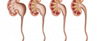
nephrectomy is most often performed , the indications for which are tumors (Wilms tumor) and malformations of the kidneys, “closed” hydronephrosis, multicystic disease (see pictures below), manifested by the presence of a tumor-like formation determined by palpation of the abdomen, dysfunction kidneys, as well as displacement and dysfunction of the abdominal organs.
Much less often, a kidney has to be removed if it ruptures as a result of a birth injury.
An important human organ is the kidney.
Acting as a kind of filter, the kidneys purify body fluid and return some of it to the bloodstream after purification. The remaining fluid, entering the ureter from the kidneys, is removed from the body. Thus, a person should be grateful to the kidneys for eliminating all harmful and toxic substances from the body.
Filtration occurs in two kidneys, since they are a paired organ.
Each kidney consists of small renal calyces. Urine collects in them and passes through their walls, filtering. The kidneys are connected to the ureters by the renal pelvis. Wider in the upper part, connected to the kidney, they become significantly narrower in the lower part, and outwardly somewhat resemble a funnel.
Anatomy and physiology
The peculiarities of the kidneys of a newborn are that the maturation of the organ is not complete by the time it leaves the mother’s abdomen. Considering organs from the point of view of morphology and functionality, they cannot be classified as full-fledged. In a newborn and a small child, the size of the organ is relatively larger compared to adults. In adults this is approximately one two-hundredth of the body part, while in a child it is one hundredth. They are located below the iliac crest. At the first stages of a child’s life, the kidney has a lobular structure, the fat capsule is almost invisible.
The renal cortex is also underdeveloped. Therefore, the medulla pyramids almost touch the capsule. However, not everything is different between children and adults. Thus, the number of nephrons exhibited by each kidney in both adults and children is approximately one million, but their size is smaller. The basement membrane has a high columnar epithelium, which reduces the filtration surface and increases resistance. Children's tubules are usually much narrower and shorter, but the distance between the descending and ascending bends is greater. The tubular epithelium, collection tubes, and loops of Henle are not yet fully differentiated.
Typically, the completion of kidney maturation from a morphological point of view is observed by the age when children go to school. In children at an earlier age, the location of the renal pelvis is mainly intrarenal, and with the development of muscle and elastic tissues, it is rather weak. The peculiarity of children's kidneys is that they work in a close connection of the lymphatic renal vessels with the same intestinal vessels. This poses some danger, since intestinal infections can quickly spread to the kidneys, which provokes the development of pyelonephritis.
Renal pelvis in newborns
Today, it is possible to timely determine the presence of the disease in utero thanks to ultrasound examinations (ultrasound). At the 17th week of pregnancy and later, thanks to this study, it is possible to determine the size of the baby’s renal pelvis and correlate them with the indicators recognized as normal.
Normal size of the renal pelvis:
- Pregnancy up to the 32nd week – from 4 to 5 mm;
- Pregnancy from the 32nd to the 36th week - from 5 to 6 mm;
- Newborn – no more than 7-8 mm.
If an ultrasound shows that the size of the pelvis is greater than the specified values, it is stated that there is an enlarged pelvis. In this case, an increase to 10 mm requires monitoring the dynamics. And if the size of the pelvis is increased beyond 10 mm, specialist intervention is required.
Detection of pathology in childhood
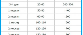
The kidneys and urinary system are formed in the fetus at 6-7 weeks of pregnancy. Enlargement of the left or right pelvis can be determined already during the second planned ultrasound (at 18-20 weeks). After birth, a control examination of the baby is carried out and the diagnosis is confirmed. In infancy, the size of the renal structures is monitored every 3 months. In addition to the instrumental technique, for children over one year of age, radiography is prescribed, and the composition of blood and urine is analyzed in the laboratory. When palpating the abdomen, the doctor detects megacystis (enlarged bladder).
Laboratory research
To diagnose pyeloectasia, laboratory tests are first prescribed to confirm the presence of abnormalities in the functioning of the urinary system. If the renal pelvis in a child is enlarged, changes in the composition of urine and blood are observed, and the volume of urine excreted is disrupted. Therefore, the doctor gives a referral for the following examination:
- General blood analysis. During the inflammatory process, the level of leukocytes increases.
- General urine analysis. There is a change in the composition of urine: the presence of red blood cell protein, an increased level of white blood cells. The urine becomes cloudy, with a white sediment.
- Urine collection according to Zimnitsky. It is carried out to study the functionality of the kidneys. Most often, oliguria (lack of urine) and hypersthenuria (increased density) are diagnosed with pyeloectasia.
- Study of urine according to Nechiporenko. An average portion of urine is taken for analysis. In the presence of kidney pathology, there is an increased level of leukocytes, red blood cells and casts.
- Bacteriological culture of urine. Necessary for identifying infectious pathogens and determining its sensitivity to antibiotics.
After laboratory examination, differential diagnosis is carried out. Changes in urine and blood may be signs of pyelonephritis, hydronephrosis, cystitis, and urolithiasis.
How does an enlarged renal pelvis affect the body?
For specialists, an enlarged renal pelvis in children is evidence that there are disturbances in the outflow of urine into the ureter. Urine accumulates in the pelvis, increasing its volume. And although dilation of the renal pelvis is not an independent disease, it has serious consequences. Because of this, without appropriate intervention and correction, serious changes in the body may occur:
- Tissues atrophy;
- Sclerosis develops;
- Kidney failure develops.
As we see, even the absence of painful manifestations with a slight increase in the renal pelvis can lead to disruptions in the functioning of such a serious organ as the kidneys.
Functionality of the organ
Already the first minutes of a child’s life become functional for the kidneys. The renal blood flow of the newborn becomes more pronounced, and the kidney begins to perform homeostatic functions. In addition, there are seven more functions for which this bean-shaped internal organ is responsible.
Distillation of plasma by the kidneys in children is much less than in adults. However, after a year, the level becomes approximately the same, if we talk about absolute and relative values. As a filter, the kidneys of a newborn work poorly, since the cubic epithelium is small in size and has low hydrostatic pressure.
Compared to adults, the volume of ultrafiltrate in children is four times less. However, during the development and growth of the child, the glomerular filtration rate increases. There is a convergence with the level of an adult by the end of the second year of life.
Newborns have lower rates of tubular reabsorption of substances of different nature, therefore more amino acids, phosphates and other substances are excreted in the urine. However, in comparison with adults, newborns have almost no differences in amino acid concentration in the blood plasma. The formation of the reabsorption system is carried out gradually, which leads to a tenfold increase.
With glucose things are different. In the fetus, the formation of the glucose reabsorption system occurs simultaneously with glomerular filtration, so glucose is stored, representing a substrate that is important from an energetic point of view.
Newborns are characterized by fairly intense reabsorption of sodium ions. In adults, with a large amount of sodium chloride, its absorption is inhibited, while in children its ions are intensively reabsorbed. This is why newborns experience swelling much more often.
The reason for the enlargement of the renal pelvis in a newborn
Currently, various causes of pelvis disease have been identified.
Congenital enlargement in most cases is inherited. Individual characteristics of intrauterine development can also lead to pelvic enlargement of the kidneys (formation of a narrowed ureter, bending it).
Complications that arose during pregnancy due to uncontrolled use of medications, disruption of a healthy lifestyle by the mother - all these are also reasons why the renal pelvis is dilated.
The reasons for the acquired enlargement of the pelvis are also complemented by complications after kidney diseases suffered by the child, and an infection that has entered the body.
Stages of the disease
Congenital or acquired pathology, dilatation of the renal pelvis can occur on each of the two kidneys. Bilateral expansion is also possible, when both pelvis are enlarged.
The enlargement of the renal pelvis in newborns occurs gradually. This allows us to distinguish the following stages of disease development
Initial, easy stage
A slight enlargement of the pelvis did not change the functioning of the kidneys. The baby does not experience any unpleasant sensations, there are no pronounced symptoms of pathology. Its presence can only be determined by ultrasound examination.
Average
At the second, middle, stage, with a significant expansion of the pelvis, an enlargement of the kidney is also noticeable. The outer tissue of the kidney is damaged, its activity has decreased by 30-40%. When urinating, the baby becomes restless and cries, which indicates certain painful sensations. A minimal amount of blood elements may appear in the urine.
Third degree - severe
It differs from the second by an increase in the characteristic signs of the disease. The renal pelvis is dilated even more, as is the child’s kidney. The destruction of the external tissues of the kidney increases. The amount of urine produced decreases. The pain experienced by the newborn intensifies. Possible increase in body temperature.
Treatment
Having received information about what dilation of the renal pelvis is in an infant, we will find out what methods are used to treat the disease.
To determine the condition of the kidneys and ureters, newborns and children under 2 years of age should be under the close attention of parents and specialists.
Clarification of the diagnosis requires special research. An X-ray will give an accurate answer to the question of whether the baby’s renal pelvis is enlarged and how large this enlargement is.
If an enlargement of the renal pelvis is detected in a newborn, the child will be placed under special registration. Regular additional studies and tests, observations of the doctor and parents will help specialists determine methods and methods of further treatment.
If there is no abnormal enlargement of the renal pelvis, the child does not experience pain, and there are no pronounced symptoms of the disease, then during the first two years no intervention in the development of the body is required. The renal pelvis system is in the process of formation, and often the child’s body, enlarging the pelvis for various reasons, then copes with the disease on its own.
The process is not ignored; the child must be under constant medical supervision. Regular urine tests and ultrasound examinations help track the dynamics.
During the first and second stages of enlargement of the pelvis, very often the enlargement of the organ goes away on its own and the problem disappears.
In the event of a significant increase in the pelvis or a rapid change in its size, the child’s body needs help.
Modern medicine uses conservative and surgical methods to treat the disease.
In conservative treatment, medications based on medicinal herbs are used, and special physiotherapy procedures are prescribed. This will make it easier for the child to urinate and stop urine from stagnating in the pelvis.
Surgical intervention is prescribed in the most severe cases of the disease, if the size of the renal pelvis has not recovered on its own, and conservative treatment has not given the expected results. The surgeon, using the laparoscopy method, makes the necessary correction to remove obstacles to the passage of urine.
Kidney nephrectomy
Indications for nephrectomy, tumors, malformations (hydronephrosis, multicystic disease) and kidney injury.
Contraindications: bilateral damage (polycystic disease) or lack of function of the opposite kidney, damage to a congenital solitary kidney.
The position for nephrectomy is on the side opposite the side of the lesion, with a bolster under the lumbar region.
Access, lumbotomy for preoperative diagnosis of kidney damage. In cases where the abdominal location of the tumor was suspected and the operation began with laparotomy, the wound of the abdominal wall is expanded and the kidney is removed through it.
Nephrectomy: progress of the operation
After dissecting the skin, subcutaneous tissue and thoracolumbar fascia, the external and internal oblique muscles are divided. Then the fibers of the transverse muscle are moved apart and the transversalis fascia is dissected, after which the perinephric tissue is exposed. The quadratus lumborum muscle is retracted to the spine, and the edges of the wound are spread apart, being careful not to damage the transitional fold of the peritoneum with the hooks.
It is better to begin isolating the kidney during nephrectomy from the upper pole, while trying not to damage the thin diaphragm and parietal peritoneum covering the kidney from the abdominal cavity. In this case, the found vessels going to the pole of the kidney from the diaphragm are ligated and crossed. Going around the kidney along the dorsal-external curvature, peel off the tissue, approach the lower pole of the kidney and free it.
Then, going around the upper pole, the kidney is separated from the adrenal gland.
After mobilization, the kidneys begin to isolate the vessels of the pedicle. The renal arteries and veins are sequentially isolated, each of which is ligated with 3 silk (silk No. 4) ligatures: 2 proximally and 1 at the renal hilum.
The vessel is crossed between them.
After this, the kidney is finally separated from the adrenal gland, which is immediately covered with a damp cloth. Then they begin to isolate the ureter. It is pulled up and, using a tampon, peeled off from the peritoneum, isolated to the point where it enters the bladder.
The ureter, tied with catgut (3 ligatures), is crossed, the stump is lubricated with iodine solution.
Produce thorough hemostasis in the wound. After nephrectomy, a drainage tube is placed to the renal bed.
The wound is sutured in layers.
Technical features of kidney nephrectomy
The technical features of kidney nephrectomy are largely determined by the nature of the pathological process for which the operation is performed.
During nephrectomy for a traumatic rupture of the organ, imbibition of the surrounding tissues by coagulated blood can create certain difficulties in isolating the renal pedicle and the vessels leading to the adrenal gland. This requires particularly careful and painstaking removal of clotted blood and drying of the wound. In case of ongoing bleeding, hemostatic clamps should be applied only to a clearly visible vessel pressed with a tupper, so as not to damage the duodenum or cecum on the right through the peritoneum, or the tissue of the pancreas and spleen on the left.
In case of hydronephrosis, when the large size of the altered organ does not allow the kidney to be “dislocated” into the wound during nephrectomy, it is advisable to apply a purse-string suture to the wall of the pelvis and puncture the cavity, evacuating the contents as much as possible, and then, after removing the needle and tightening the purse-string suture, continue isolating the kidney.
Disease prevention
Timely intervention on the pathology that appears in the child will preserve the healthy functioning of the kidneys and protect them from infections and other diseases.
Young parents, having learned about the diagnosis of “dilated renal pelvis,” should not despair! They are required to become even more attentive to all the life processes of the baby, and receive the necessary consultations from a pediatrician and pediatric urologist.
To prevent the disease, parents should ensure:
- Ultrasound of the kidneys and urinary tract - the first month of life.
- In case of pathology - regular urine testing and consultation with a doctor.
- If the pelvis is slightly dilated, an ultrasound scan is performed once every three months.
The joint care of loving parents and professional specialists, as well as timely medical assistance will help maintain the health of the child!