A Mongolian or Mongoloid spot in a newborn is a congenital nevus that becomes bluish in color and looks like a hematoma. It is a type of birthmark and may disappear on its own over time. Let's take a closer look at the reasons for the appearance of neoplasms in children.
What is it and features
The Mongolian spot in children is a congenital pathology, and is an accumulation of melanin in the middle layers of the skin. This type of birthmarks acquired this name not because of Mongolia itself, but because of the nature of its manifestation in children of the Mongoloid races. More than 80% of newborns in China, Korea, Vietnam, Japan, Indonesia, India, and Chukotka are born with blue spots on their bodies. Slightly less often, such manifestations on the body are found in children of the Negroid race and among the population of the Earth, distinguished by a dark or dark skin tone. Only more than 1% of white-skinned children are born with this pathology in European countries.
This phenomenon cannot be called a disease; the stain does not threaten the health and life of the child, and is not accompanied by any symptoms, unpleasant sensations or discomfort. Nevus is only a cosmetic problem that goes away on its own by 2-4 years of age.
The German anthropologist Egon von Eickstedt was interested in Mongoloid spots on the body and noted that they are hereditary. Some nationalities consider this manifestation to be an imprint of higher powers and attach divine significance to the marks, while Brazilians and residents of northwestern South America carefully hide the blue marks and consider them something of a shame or an extreme nuisance. Eskimos, Mongols, Buryats and Polynesians regard the blue sign on the body of a newborn as a favor towards man from Nature itself and Heaven. The larger the spot, the more blessed the child will be. This phenomenon on the skin began to be called sacred. Less religious peoples nicknamed the gray-blue pigmentation “coccygeal spot” due to its frequent location in the sacral spine.
We recommend reading
- What can cause nipples to darken?
- How to whiten dark spots under arms
- All kinds of methods to get rid of brown spots on the face
The famous anthropologist and physician Erwin Beltz was inclined to think that education among representatives of the Caucasian race can only happen when there is racial admixture in the family.
Mechanism of color spot formation
To find out the origins of pathology, you need to understand what it is and how the process of skin pigmentation occurs. Color is influenced by melanins - pigments of complex structure and chemical composition. They are divided into
- Pheomelanins have a red tint and impart it to organs: lips, nipples, genitals. Hair saturated with pheomelanin pigment will have a red color.
- Neuromelanin is found in brain tissue. Its purpose has not been revealed.
- Eumelanins are predominantly brownish or black in color.
Melanins create our appearance, saturating it with different colors. They are also found in other parts of the body. This pigment is produced by specialized cells - melanocytes.
The human skin consists of two layers:
- upper (epidermis, consisting of squamous epithelium);
- internal (dermis - a porous area created by connective collagen tissue).
Skin pigmentation occurs in the epidermis. Melanocytes produce the European melanin pigment when exposed to ultraviolet rays. The remaining races tirelessly produce melanin with melanocytes, creating the color of the skin. The dependence of color turned out to be not in the number of melanocyte cells, but in their activity. If there are not enough effective melanocytes to migrate between the skin layers during the development of the embryo, then a failure occurs, and a certain number of them remain in the dermis, creating a gray-blue tint in places of accumulation.
These are the causes of Mongolian spots in newborns.
Causes and manifestations in a newborn
Medical experts explain the formation of a characteristic bluish tint on the skin of a newborn baby as an unfinished process in the migration of cells responsible for the synthesis of melanin during the development of the embryo. These cells are called melanocytes and they produce natural pigment located in the basal layer of the epidermis. The melanin produced by these cells determines the color of a person's skin. In babies in the womb, during their development, melanocytes migrate to the upper layers of the dermis from the ectoderm. In some newborns, this process is incomplete, and the melanocytes remain in the deeper layer of the integument, and the synthesized pigment instead of a brown tint appears bluish. The reason for the formation of a Mongoloid nevus in a child is a deeper location of melanocytes than expected.
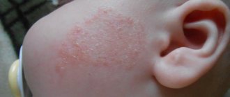
At birth, this pigmentation causes uninformed parents to panic and worry about the baby’s health. This is not surprising, especially when the manifestations on the body are extensive and the color of the lesions is rich. Pathology is expressed differently in different cases. Sometimes the spots are the size of a coin and are single, in other children they are large and occupy a large area, sometimes an entire part of the body. The color of the problem area can vary from bluish-lilac and gray-blue to dark blue, almost black. Among Indians, such spots are often very dark and have a green tint.
The characteristic of the spots is that they have a uniform color throughout the pigmented area. They do not hurt, do not itch, and do not rise above the level of the rest of the integument. The shape of the formations is often irregular; small manifestations on the skin can be round or oval.
Based on the observations of specialists and parents of children with pigmented skin, one more feature of Mongoloid nevi can be identified: provided that they are represented by several spots, hyperpigmented areas can change their location and merge into one gray-blue area. So the affected area may be a fairly large part of the skin: the entire back, arm, buttocks, leg.
A characteristic feature is the fading of pigmentation as the child ages. The bluish areas of the body gradually acquire a normal color and by 3-4 years the spots disappear without a trace. In rare cases, the pathology does not go away on its own, and in 5% of newborns with blue nevi, accumulations of melanin in the middle layers of the skin remain in adulthood.
We recommend reading
- Pigment spots in newborns: causes, treatment
- Causes and symptoms of white spots around the eyes
- Features of white spots in children: how to get rid of them
Appearance
The dark mark is a congenital nevus. In most cases, the Mongoloid spot in a newborn has a blue-gray color, reminiscent of a bruise. Sometimes these spots are blue-black or blue-brown. A distinctive feature of these particular spots is considered to be uniform coloring throughout the entire area with altered pigmentation.
The shape of the spot can be completely different, mostly irregular in shape. Sizes also do not have standards - they range from specks no larger than a coin to large spots covering the entire back.
The Mongoloid spot in a newborn is most often concentrated on the lower back or sacrum. But other places of manifestation are also quite likely: spots are known to appear on the legs, back, forearms and even hands. Even migrating spots are very rare, gradually moving, for example, from the buttocks to the lower back and back.
Most often, the spot is present in a single copy, but there are also manifestations of multiple marks.
Immediately after birth, the “blobs” darken, but over time they become paler and smaller. In almost all children, by the age of 5, the skin acquires a uniform color. Rarely, marks can be found in adolescents. Mongoloid spots remain in an adult only if there were a lot of them in childhood, and in atypical places.
Localization Features
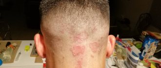
Mongoloid spots are also called coccygeal spots. The second name is associated with their frequent location. Often the pigment island in a newborn is located on the butt, in the tailbone area and on the buttocks, as well as on the lower back and hips. With extensive damage, pigmentation can occupy the entire back, including the neck and head of the child. Often the site of localization is the perineum and lower extremities, especially the back of the legs. Less commonly, islands are found on the arms, abdomen, and face.
Attitude
The Mongoloid spot, a photo of which accompanies this article, has different meanings among different peoples. For example, in Brazil they consider it a shame to have such marks; parents carefully hide this fact even from their closest relatives, not to mention strangers. In addition, the color of the spot among the inhabitants of Brazil is close to greenish, therefore, if a nevus is suddenly discovered in an adult, he will be teased as “green-backed.”
For most peoples, a stain is a “slap from Buddha”, “a kiss from God”. It is believed that a child with such a mark will be happy, since God (Buddha, Allah) is looking after him. And, of course, this is another opportunity to make sure that the child is a representative of a certain people.
Treatment in newborns
Having discovered bluish flat formations on the child’s body, parents should consult a dermatologist, despite their benign quality and asymptomatic occurrence.
There are other types of nevi, which are similar in appearance to Mongoloid pigmentation, and only a doctor will be able to differentiate these manifestations from other, more dangerous phenomena. It is necessary to distinguish Mongolian spots from nevus of Ota, blue and pigmented giant birthmarks.
Diagnostics is necessary to exclude formations that can degenerate into melanoma - a cancer of the skin.
To diagnose pathology, a dermatologist performs dermatoscopy and siascopic examination. These procedures are non-invasive and allow you to determine the nature of the formation. A histological examination of the neoplasm cells may also be prescribed, which allows one to determine the location of the melanocytes. Normally, they should be located in the epidermis - the upper layer of the skin. In the presence of a Mongolian nevus, cells that produce melanin are located in the dermis. The detection of melanocytes in the lower layer of skin will mean that the formation is Mongolian. When such a diagnosis is made, treatment of pigmentation is not required.
How to deal with uncharacteristic pigmentation of the epidermis?
First you need to show your child to a dermatologist. The doctor will examine the patient, conduct a series of tests and make a diagnosis. Mongoloid neoplasm goes away on its own. By the age of 20, it disappears from the body completely. This often happens in early childhood, namely at the age of 4-6 years. So no need to worry. It is recommended to remove Nevus Ota with laser in childhood. This will eliminate the risk of developing melanoma. The same applies to Ito-type education.
Nevus Ota is recommended to be removed in childhood
The doctor chooses the method of getting rid of the tumor. It could be:
- laser removal;
- use of radio waves;
- surgical intervention;
- restoration of normal pigmentation using liquid nitrogen.
The older the patient, the more likely complications are to occur. Especially if the nevus of Ota is to be removed. After treatment, a number of restorative procedures are prescribed. They help the epidermis regenerate and prevent melanocytes from being reactivated under the influence of a foreign gene. Patients who have had a combined nevus removed should register with a dermatologist so that the doctor can monitor changes in the skin. In case of relapse, the spot is eliminated again so that it does not develop into a cancerous tumor.
Forecast
The presence of a gray-blue spot in a child, with a correct diagnosis, should not worry parents. In medical practice, not a single case of degeneration of a Mongoloid nevus into a malignant formation has been recorded.
When the baby is born, the spot may have a rich, characteristic shade, but over time the island will lighten until it ceases to be noticeable at all. In 90% of cases, the mole disappears by 2 years of age, in 3% by 4-5 years of age, in 2% by 13-15 years of age, and only about 5% of people with this pathology remain with it for life. The latter occurs in patients who have extensive damage to the body with pigmentation with a rich color.
Prevention
Since God's Mark is not a disease, there is no cure for it. The prognosis for such a nevus is positive. During the entire period of observation of these spots, not a single case of its degeneration into melanoma was recorded. For this reason, there is no need for medical supervision.
In most cases, the spot disappears on its own by age five. But even in those rare cases when it remains for life, it does not have any effect on the health or functions of the body.
Diagnostics
If you find an unknown spot on your child’s skin, you should consult a dermatologist. The doctor will conduct a special examination to make sure that these are not pathological pigmented nevi, since some of their varieties can be melanoma-dangerous. If one of these options is detected, it is necessary to be constantly monitored by a dermatologist and oncologist.
To distinguish the Mongoloid spot from other types of nevus, siacopy and dermatoscopy are performed. If the diagnosis needs clarification, the doctor may prescribe a biopsy of the pigmented area.