Epidermolysis bullosa is divided into four main types, differing in the level of skin at which the blisters form:
- epidermolysis bullosa simplex (EBS) - in the upper layers of the epidermis;
- borderline epidermolysis bullosa (BEB) - at the level of the light plate (lamina lucida);
- dystrophic epidermolysis bullosa (DEB) - in the upper part of the papillary dermis, below the lamina densa;
- Kindler's syndrome - different levels of blistering.
Each type is characterized by its own severity of manifestations and different combinations of mutations in different genes. The types of mutations also determine the nature of inheritance of the disease: autosomal dominant or autosomal recessive. Currently, 18 genes have been identified, mutations in which are associated with various subtypes of EB.
| Main type of BE | Main subtypes of EB | Target proteins |
| Simple BE (SBE) | Suprabasal PBE | plakophilin-1; desmoplactin; maybe others |
| Basal PBE | α6β4-integrin | |
| Borderline BE (BBE) | PoBE, Herlitz subtype | laminin-332 (laminin-5) |
| PoBE, others | laminin-332; collagen type XVII; α6β4-integrin | |
| Dystrophic EB (DEB) | dominant DBE | collagen type VII |
| recessive DBE | collagen type VII | |
| Kindler syndrome | — | kindlin-1 |
Epidermolysis bullosa: definition
Pathology is a group of skin diseases, hereditary or acquired (less common), that cause blisters to appear on the skin. Blisters can form in response to minor injuries (bruise, friction, burn). For this reason, the disease was given another name - “mechanobullous disease”.
In most cases, epidermolysis bullosa is inherited. Appears in infancy or early childhood. It usually affects the skin of the lower and upper extremities. In some children, signs and symptoms of the disease do not appear until adolescence.
For reference: “Butterfly babies” is the name given to babies who are born with this disease, since their skin is considered as delicate and fragile as the wings of a butterfly.
Mechanobullous disease (epidermolysis bullosa) cannot be completely cured due to the lack of special medications. But some of its forms (localized) improve as the child grows older.
Alleviation of the disease occurs by eliminating symptoms such as itching, preventing pain and healing wounds. Severe forms of the disease, when blisters appear inside the body, such as on the lining of the mouth or intestinal walls, often cause serious complications and death.
Clinical manifestations of BE
Epidermolysis bullosa manifests itself already during childbirth: the baby’s skin is injured when he passes through the birth canal. The nose, chin and heels are usually affected. In rare cases, the disease makes itself felt at 1-6 months of life. With age, depending on the type of EB, new symptoms of the disease may appear.
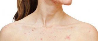
The main clinical manifestations of epidermolysis bullosa are blisters on the skin that appear at sites of friction, bruise, pressure, with increased body temperature, the environment, or spontaneously. Blistering can also occur on the mucous membranes of any organ. Most often the mucous membrane of the oral cavity, esophagus, intestines, genitourinary system, and eye mucosa are affected.
Why do blisters form?
Human skin consists of two layers: the outer layer is the epidermis, and the layer underneath is the dermis. In people with healthy skin, between these two layers there are anchor proteins that prevent the epidermis and dermis from moving independently of each other.
People born with epidermolysis bullosa (EB) lack the bonding proteins that cause the skin to become extremely fragile. Even a slight mechanical effect (friction, pressure or injury) contributes to the separation of its layers. As a result, blisters and painful wounds form on the body.
Note! Skin ulcers in patients with EB are similar to third-degree burns. In addition, people suffering from epidermolysis bullosa are at increased risk of developing cancer.
Causes of the disease
Epidermolysis bullosa is caused by defective genes. A child may inherit them from one or both parents.
There are cases when neither the mother nor the father of the child had EB, but the baby was born with signs of the disease. This happens when both parents are hidden “carriers” of mutation genes.
Although there are often cases when a defect in the genes of a healthy person occurs spontaneously, then the pathology is called acquired.
How is EB inherited?
All genes enter the human body in pairs. A child receives half of its genes from its mother and the other half from its father.
However, the genetic conditions of epidermolysis bullosa and the mutations that cause this disease can be transmitted from generation to generation in two ways:
- Autosomal dominant. It only takes one defective gene in one of the parents for the newborn to develop symptoms of a certain genetic condition, in this case epidermolysis bullosa. The disease is then diagnosed in each generation. The risk of inheritance is 50%.
- Autosomal recessive. Husband and wife have genes with the same defect. However, they do not have any symptoms of EB, since they are only carriers of mutated genes. The probability that children, like their parents, will become carriers of a latent defect is 50%. The chance of having babies without the mutation is 25%, and the chance of having a baby with epidermolysis bullosa is 25%.
For reference: Epidermolysis bullosa affects both men and women with equal frequency. Pathology occurs immediately at birth or shortly thereafter.
Classification of the disease
Acquired epidermolysis bullosa in children and adults can occur in several ways.
Specialists in the field of dermatology highlight:
- Epidermolysis bullosa simplex. It differs in that it is inherited in a dominant manner and is caused by the presence of mutations in the KRT5 and KRT14 genes. Geneticists note that an incorrect structure in these genes is observed only in 75% of cases of pathology development. Damage affects proteins such as keratin 5 and keratin 14, alpha 6 and beta 4 integrin, and plectin. It has 12 subtypes, among which the most common are Weber-Cockayne, Koebner and Dowling-Meara syndromes. The diagnosis rate is 1 in 100 thousand.
- Borderline. It has a recessive type of inheritance and is caused by a violation of the structure of the LAMB3 and LAMA3 genes and some other segments. It is characterized by a more severe course and involvement of the protein laminin 332, collagen 17, alpha-6 and beta-4 integrin. The peculiarity is that this variety has 2 subtypes and 6 independent forms. This diagnosis is given to 1 person per 500 thousand population.
- Recessive dystrophic epidermolysis bullosa. It can be inherited both recessively and dominantly. Structural lesions develop in the COL7A1 gene, and the target protein is collagen 7. There are several clinical types, the most severe of which is Allopo-Siemens syndrome.
- Kindler's syndrome or mixed epidermolysis bullosa. Currently, among clinicians it is considered one of the rarest and least studied types of the disease. In such cases, only one protein acts as a target—kindlin 1.
Such mechanisms of the course of the disease affect the life expectancy of epidermolysis bullosa, which is purely individual for each patient.
The simple form of the disease has its own classification and happens:
- localized;
- generalized.
According to the etiological factor, they distinguish:
- congenital epidermolysis bullosa - a defect in genes can occur at the stage of conception or during pregnancy;
- acquired epidermolysis bullosa - develops according to the type of autoimmune pathologies (the causes of the disease have not been established by specialists).
Methods for diagnosing the disease
If a doctor, when examining your skin, suspects you or your baby has epidermolysis bullosa, then in order to confirm the diagnosis, he will definitely prescribe the following tests for you:
- Skin biopsy for immunofluorescence mapping. A small sample of the affected skin is excised and examined to determine the extent of the damage. This accurate and reliable test allows not only to diagnose skin pathology, but also to obtain information about the characteristics of its course, as well as predict the result.
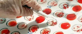
- Genetic testing. It is used to confirm the diagnosis because most often epidermolysis bullosa is inherited. For this purpose, blood from a vein is given for laboratory analysis (in a small child, blood is taken from the heel).
Attention! Prenatal testing (blood sampling from a vein) and consultation with a geneticist are prescribed for pregnant women who have a family history of epidermolysis bullosa, which allows for timely detection of pathology in the fetus.
In the following video, a geneticist will talk about what specialists can do to prevent the birth of a child with a hereditary disease:
What material is taken for research?
The subject of research in the laboratory to establish a genetic defect is the blood of a patient or a healthy carrier of a pathological gene (healthy parents of a sick child). For the initial examination, 2 ml of blood is sufficient (5 ml from the parents of a sick child). The shelf life of blood in a special tube is 2 weeks in the refrigerator, during which time the tube is turned over several times. Blood in a test tube is not the only possible object of study. You can also send a blood stain on special blotting paper for testing. In this case, there should be several spots (2-3), and the diameter of each spot should be at least 2 cm. Blotting paper soaked in blood is dried in air, placed in an envelope and sent by mail. Biological material can be taken for research directly in the laboratory.
For prenatal diagnosis of the presence of the disease in the fetus, chorionic villi (fetal membrane) are taken. Taking chorionic villi for examination is possible in several Moscow institutions (probably in other cities too), the Center for Molecular Genetics has a particularly close connection with maternity hospital No. 27 (tel. 450-47-83), where you should contact in the 9-11th week pregnancy (maximum up to the 12th), having previously agreed upon the receipt of the material in the laboratory. The material is taken on an outpatient basis (that is, there is no need to go to the hospital) under local anesthesia (anesthetic injection). A few hours after the procedure you can go home. The risk of spontaneous abortion during the study is minimal.
Treatment
It is impossible to completely get rid of the symptoms of epidermolysis bullosa, since there is no specific treatment. Doctors try to alleviate the condition of such patients by:
- Preventing the development of complications of the disease is the main task to prevent the growth of blisters and preserve their crusts. For this purpose, atraumatic non-adhesive materials are applied to the affected areas, then a bandage is applied in several layers. This approach helps prevent the appearance of ulcers and heal wounds faster.
- Opening the blisters with a sterile needle and treating them with antiseptics (chlorhexidine bigluconate solution 0.05%, 0.1%, 0.5%).
- Applying heliomycin ointment to the affected areas of the skin.
If epidermolysis bullosa has taken a complex form, then the patient is prescribed Prednisolone. For preventive purposes, irradiation of the skin with ultraviolet rays is attributed.
Below is a video for parents of children with epidermolysis bullosa. In it, a dermatologist talks about the features of the pathology and gives advice on improving the quality of life of sick children.
Question answer
What are the signs that your current treatment for epidermolysis bullosa is working?
The main signs of improvement in the condition of a patient with BE:
- The skin affected by the disease not only stops growing, but also begins to decrease in size.
- New blisters and pimples no longer appear on the skin.
- The patient's well-being gradually returns to normal.
How can you prevent skin injury?
Wear only clothes made of soft natural fabric and without rough seams (you can turn them inside out). Shoes should be soft, with rubber soles.
What should hygiene be like for epidermolysis bullosa?
The water temperature when swimming is 37-38°C. The duration of water procedures can be 30 minutes (longer is possible so that the bandages come off the skin more easily). With a cosmetic cleanser, you should only wash those areas of the skin that need daily hygiene. For this purpose, it is recommended to use special creams or shower gels that contain oils (moisturizing gel or La Roche-Posay cream).
Is it possible to add sea salt to the bath to dry out blisters?
This is not recommended. Since the skin of patients with epidermolysis bullosa is very fragile and has wounds, salt will only cause pain during bathing.
How is a genetic defect determined?
There is a special file of genetic defects found in each hereditary disease - OMIM. To narrow the search, with EB it is necessary to know the clinical diagnosis; it is established by a dermatologist, but the dermatologist cannot know what specific genetic defect a given patient has, because This diagnosis may correspond to several different breakdowns. The file contains an indication of the pathological gene and the most common defects in its various sections (exons). Thus, in recessive dystrophic EB there are 118 such areas. Some closely located areas (from 3 to 5) can be combined into one view. But even then you will need to view 44 fragments.
What to remember:
- Epidermolysis bullosa is a rare but life-threatening disease that most often appears in a person from birth.
- Pathology is inherited, but can also be acquired.
- No form of epidermolysis bullosa can be cured, since no special medications have yet been developed.
- Treatment is based on preventing complications of the pathology and taking timely preventive measures (ultraviolet irradiation).
- If a pregnant woman has had cases of epidermolysis bullosa in her family, she should definitely undergo consultation with a geneticist and prenatal testing.
Where to contact
Center for Molecular Genetics (DNA Diagnostics)
Address: 115478 Moscow, st. Moskvorechye, 1. In the building of the Medical Genetic Research Center on the 1st floor, room 116, DNA Diagnostics Laboratory, Laboratory: (495) 504-31-66, General Director Professor Polyakov Alexander Vladimirovich: (495) 727-95-02, fax, e- mail https://www.dnalab.ru/
Maternity hospital No. 27, Moscow (tel. 450-47-83)
Send the material by mail or bring it to the laboratory: Moscow, st. Moskvorechye, 1 (building of the Medical Genetic Research Center of the Russian Academy of Medical Sciences), 1st floor, room. 116 from 9.30 to 15.00. Before conducting the study, you need to call the laboratory, state the purpose of the study, receive an invoice and pay for it. If the material is sent by mail, the following information is attached to it: full name of the person in respect of whom the research is being conducted, the address to which the conclusion should be sent, a copy of the paid receipt, the purpose of the research.
Prof., Dr. med. Science Albanova Vera Igorevna