With the onset of conception, every woman is curious to know what exactly happens to the baby at this stage of pregnancy. Some mothers study in advance literature that describes in detail the development of the fetus by week of pregnancy, and after fertilization they compare their feelings with those described. Sometimes such curiosity promptly notices unusual signs and prevents the development of dangerous complications or irreversible consequences. Therefore, it would be useful for every mother to know the peculiarities of pregnancy development.
Happy moment of waiting for the baby to be born
How is gestational age calculated?
Gestational age is the main criterion by which the correct development of the baby in the womb is determined.
There are 2 stages of pregnancy:
- embryonic (true),
- obstetric.
The embryonic period starts from the moment of fertilization.
Since it is not always possible to establish the exact date of conception, in obstetrics it is customary to consider the period of pregnancy from the beginning of the last menstruation - the obstetric period.
According to the obstetric period, a physiological (normal) pregnancy lasts on average 9 calendar months, 10 lunar months (in a lunar month - 28 days), 40 weeks or 280 days.
Considering that the usual duration of a woman’s menstrual cycle is 28 days, and ovulation of a mature egg occurs on days 13-14, fertilization can occur 2 weeks from the start of menstruation. This will be considered the embryonic period of pregnancy. Thus, the embryonic period is 2 weeks shorter than the obstetric period and lasts approximately 38 weeks. Such calculations are correct only for women with a stable cycle and the absence of hormonal disorders; in other cases, deviations in terms are possible either upward or downward.
According to statistics, the discrepancy between obstetric and embryonic periods is less than 2 weeks in 20% of women, 2-3 weeks in 45%, and more than 3 weeks in 15%.
Division of the embryonic stage
Intrauterine development is represented by several stages. They depend on the characteristics of maturation and the response of the embryo to any irritating factors. The period is divided into several stages:
- preimplantation;
- implantation;
- embryo;
- fetus.
The first stage is the most important, it begins with fertilization of the egg and ends after the blastocyst penetrates the uterine lining. This process occurs 5-6 days after the formation of the zygote.
Registration of pregnant women
According to the law, a woman has the right to free medical care, regardless of when she registered with a medical institution for pregnancy. Doctors call the optimal period for registration from 7 to 12 weeks of pregnancy.
Early registration of a pregnant woman allows:
- Timely diagnose somatic diseases and pathologies of the reproductive system, including sexually transmitted infections.
- Reduce the likelihood of early miscarriage. This is especially true for women with a history of spontaneous abortion.
- Rule out ectopic pregnancy.
How much do surrogate mothers earn?
General concept of ontogenesis
This process is an individual cyclic development characteristic of all organisms. The basis is represented by the implementation of hereditary information throughout existence. It is important to understand that the influence of external factors is of great importance.
Ontogenesis is characterized by a long historical development, special for each species. Scientists Haeckel and Muller formed a biogenetic law that can describe the relationship between historical and individual improvement.
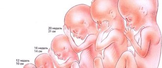
Fetal development calendar by week
The period of intrauterine development is divided into:
- embryonic or fetal – the first 8 weeks of pregnancy.
- fetal or fetal – lasts from the 9th week of pregnancy until its end.
During the embryonic period, the formation and differentiation of tissues occurs, and during the fetal period, growth and development of organs and functional systems of the fetus take place.
| Week of pregnancy | Fetal development | Changes in a woman's body |
| 1 | Preparation for possible fertilization. | |
| 2 | In the ovary, an egg matures in the dominant follicle. | |
| 3 | Conception occurs. Male (sperm) and female (ovum) reproductive cells contain half the set of chromosomes. When they merge, a zygote is formed - the first cell of a new organism with a full set of chromosomes. | During ovulation, a mature egg is released from the ovary into the fallopian tube, where it is fertilized by a sperm. |
| 4 | The zygote begins to divide and move towards the uterus due to peristalsis of the fallopian tube. This journey takes 3-5 days. A multicellular embryo enters the uterine cavity. The embryo is implanted into the uterine mucosa. The size of the embryo is 1.5 mm. | The next menstruation does not occur. The first subjective signs of pregnancy appear: weakness, drowsiness, engorgement of the mammary glands, mood swings. The pregnancy test is positive. |
| 5 | The embryo is about 2 mm, consists of 3 layers: ectoderm, mesoderm and endoderm, from which the fetal organs will be formed. The anterior and posterior poles (the future head and legs) are determined in the embryo. The body develops along the axis of symmetry - the chord. | Characteristic symptoms of early toxicosis include the appearance of nausea, vomiting in the morning, and intolerance to strong odors. |
| 6 | The embryo grows to 6-7 mm. The rudiments of arms and legs appear, the heart is already divided into chambers, pulsating, and the hemispheres of the brain are taking shape. The future placenta is laid, the circulatory network is actively developing, through which the embryo will receive the necessary nutrients and oxygen. | Signs of early toxicosis intensify. |
| 7 | Embryo weight – 1 g, height 8-11 mm. Facial features are already visible - nose, eyes, mouth. The brain is developing at a rapid pace. The umbilical cord and uteroplacental circulation are formed. | The uterus increases in size. Symptoms of toxicosis persist, accompanied by severe weakness and drowsiness. Urination becomes more frequent. |
| 8 | The embryo is the size of a bean, weighs 5 g. Facial features continue to develop, ossification of the bones of the skull, arms, and legs begins. The formation of internal organs is underway: heart, gastrointestinal tract, kidneys, bladder. The baby begins to move, but the mother does not yet feel these movements. | The state of health remains the same. |
| 9 | The baby is 35-45 mm long, weighs 10 g, the head occupies more than half the body length. The reproductive system is being formed, the adrenal glands are already synthesizing hormones, and the liver is producing new blood cells. | Signs of toxicosis still persist, but the body is already adapting to the new state and feeling better. |
| 10 | The first critical period, the embryonic period, ends. The fetus is actively structuring its organs: a septum has been formed between the chest and abdominal cavities, and the kidneys begin to produce urine. Up to 250 thousand neurons are produced in the brain every minute. | The uterus is enlarged, but there are no external signs of pregnancy yet. The symptoms of toxicosis pass. |
| 11 | At this stage (11-13 weeks), the first ultrasound is performed. The head is large relative to the body, the arms are long, and the legs are short and bent at the knees. There are already rudiments of teeth and nails. | Gastrointestinal symptoms may appear: heartburn, constipation, tendency to flatulence. |
| 12 | Weight – 20 g, length about 9 cm. The fruit actively moves its arms, legs, and fingers. The intestines fold into loops, and leukocytes appear in the blood, whose function is to protect against infections. | Due to changes in nutrition, metabolism, and hormonal levels, the figure begins to change, weight gain is 1-2 kg. |
| 13 | A new trimester of pregnancy begins. All the main organs and systems have already been formed, and then they will have to grow and develop before birth. The face becomes human-like, the mouth has the beginnings of 20 baby teeth, and the first hairs begin to grow. | The shape of the abdomen changes. Emotionally, the woman becomes calmer. |
| 14 | The weight of the fetus reaches 45 g, height 13 cm. Differentiation of the genital organs is underway: in boys the prostate is formed, in girls - the ovaries. The sucking reflex and imitation of breathing movements are triggered. The pancreas begins to produce insulin. The main coordinator of the endocrine system, the pituitary gland, comes into play. | The pregnant uterus rises 10-15 cm above the pubis. The woman can feel her upper pole. |
| 15 | Weight is about 70 g. The skin is covered with vellus hairs, which help retain heat. The baby is actively moving and making faces. A special protein appears in the blood that determines the blood group. | Pigmentation of the skin appears: on the nipples, white line of the abdomen. |
| 16 | The fruit is similar in size to an avocado. The skeletal bones are made stronger, but at the same time remain flexible, so that during childbirth the baby can pass through the birth canal. In girls, egg primordia are formed. | Normally, weight gain by this period reaches 2-3 kg. |
| 17 | The placenta supplies the baby with nutrients and oxygen and removes waste products. The weight of the fetus reaches 150 g, height 15 cm. Subcutaneous fat begins to form, sweat glands develop. | As the volume of circulating blood increases, problems with the functioning of the heart may occur: tachycardia. |
| 18 | Weight - 250 g, he already hears sounds, and soon he will be able to recognize his parents' voices among other noises. | The expectant mother feels the baby move for the first time. |
| 19 | Weight – 300 g, height – 25 cm. The anatomical structure of the fetus becomes more symmetrical: the head does not grow so quickly, and the torso and limbs continue to lengthen. A cheese-like lubricant thickly covering the entire skin surface of the baby helps regulate body temperature. The rudiments of molars are formed. | The rapidly growing uterus is located 1-2 cm below the navel. Due to stretching of the uterine ligaments, painful sensations in the lower abdomen may periodically occur. |
| 20 | This week is the middle of pregnancy. The baby weighs 350 g, actively moves his arms and legs, and can suck his fingers. Becoming more and more like his parents. | An enlarged uterus compresses the insides, and as a result, a woman may experience shortness of breath and frequent urination. |
| 21 | Weight – 400 g, height 30 cm. Most of the nutrients come through the placenta. The stomach is already capable of digesting amniotic fluid if it enters the digestive tract through swallowing. The baby's taste buds begin to function. | Body weight increases, the configuration of the figure changes. |
| 22 | By the end of the week, the weight of the fetus will reach 500 g. The skin is red, wrinkled, covered with grease. With the development of nerve receptors, the baby begins to respond to touch. The brain from 21 to 25 weeks increases 5 times, from 20 g to 100 g. | |
| 23 | Maturing brain cells take control of all life processes of the fetus: movement, sensory organs and others. A substance begins to be synthesized in the lungs that will allow them to expand and breathe after the birth of the child. | The height of the uterine fundus is 4 cm above the navel. The large volume of the uterus leads to discomfort in the spine and joints. At this time, it is recommended to wear a prenatal bandage. |
| 24 | Weight – 600 g, height – 33 cm. As the vestibular apparatus matures, the baby begins to meaningfully navigate the uterine cavity (understands where is down and where is up). | Body weight gain continues - up to 500 g per week. Pastosity of the lower extremities and swelling may appear. |
| 25 | Weight – 750 g. The bone and joint system is actively developing. Meconium is formed in the large intestine - the original feces that will be released after the birth of the child. | There is a high likelihood of developing iron deficiency anemia. If fatigue, weakness, pallor, or tachycardia appear, consult a therapist. |
| 26 | Weight – 900 g, height – 34 cm. The lungs continue to produce substances that will prevent them from sticking together after the newborn’s first breath. The baby develops periods of sleep and wakefulness. | |
| 27 | The weight of the fetus is approaching a kilogram. The pituitary gland produces growth hormone, and the thyroid gland produces hormones that regulate metabolism. | Sensory disturbances may appear in the legs: tingling, goosebumps, even cramps. |
| 28 | The baby can open and close its eyes. The iris of the eyes acquired color due to the accumulation of pigment, but this color is not final. | Body weight gain is 7-9 kg . So during pregnancy, the muscles of the venous walls also relax, this can manifest itself as varicose veins of the legs. |
| 29 | The baby is becoming stronger and more active every week. He is already showing character, which is expressed in different reactions to external stimuli: light, sound. | Some women continue to be bothered by heartburn, constipation, exacerbation of hemorrhoids, and frequent urination. |
| 30 | The baby is actively gaining weight, but movements are slowing down, due to an increase in its size. | Pregnant women feel engorgement of the mammary glands; when pressing on the nipples, colostrum is released. |
| 31 | Weight – 1600 g, height – 40 cm. Before birth, the baby will be in the uterine cavity in the fetal position (arms pressed to the chest and legs pulled up to the stomach). In boys, the testicles begin to descend into the scrotum. | At this time, signs of gestosis may appear: increased blood pressure, swelling. A pregnant woman should not lie on her back, as in this position the enlarged uterus puts pressure on the inferior vena cava, which can cause a drop in pressure. |
| 32 | This week marks the end of another critical period. All major organs, except the lungs, are able to function fully in case of premature birth. | A pregnant woman may experience increased pain in the spine, joints, and pubic symphysis. |
| 33 | Weight is about 2 kg, it is already difficult for him to move. His activity is largely influenced by his mother's nutrition, sounds, and walks. | The height of the uterine fundus is 34 cm above the level of the pubic symphysis. It becomes difficult for a pregnant woman to endure physical activity. |
| 34 | The fetus begins to prepare for birth. The original lubricant gradually disappears from the skin, lingering in large folds: behind the ears, under the arms, in the groin. | A pregnant woman may experience episodes of false contractions - the myometrial muscles are preparing for the birth process. |
| 35 | Height 47-48 cm, weight 2300-2500 g. The fetus occupies the physiologically correct position - head down. | Due to changes in the position of the heart during physical activity, shortness of breath often occurs. |
| 36 | The fetus continues to accumulate fat, which is extremely important for the child after birth to retain heat and obtain energy. The sucking reflex is well developed. | The height of the uterine fundus above the womb is 36 cm. Hormonal changes in the woman’s body continue, preparing her for childbirth. |
| 37 | Weight 2600-2800 g. The volume of subcutaneous fat reaches 15% of the total body weight of the fetus. The vellus cover on the baby's body gradually disappears. | A pregnant woman experiences signs of labor: the volume of the abdomen decreases, the fundus of the uterus descends, false contractions intensify, and a mucus plug comes out of the cervical canal. |
| 38 —40 | Normally, the baby's head has dropped into the pelvis, which corresponds to a cephalic presentation. The birth of a child at this time is considered physiological, the baby is considered full-term. | Labor can begin at any time. |
Of the 75-900 million sperm that enter the vagina during intercourse, only a few thousand will reach the uterus and only one of them will fertilize the egg.
Main stages
From a biological point of view, the main event in the entire development of individual organisms is represented by the ability to reproduce. It is the key to the existence of certain species in natural conditions. Taking into account the characteristics of reproduction, ontogenesis is divided into periods:
- pre-reproductive;
- directly reproductive;
- post-reproductive.
The first stage is the implementation of the transmitted hereditary information. It is expressed by functional and structural changes in the body. At this time, the individual shows increased sensitivity to any actions. The stage can be embryonic, metamorphotic, larval, or juvenile.
During the reproductive period, each organism manifests its main purpose, which is to procreate. As for the last stage, it is inevitable for individual development. Any living creature undergoes aging, followed by a decline in basic life functions, which ends in death.
Changes in the body of a pregnant woman
During pregnancy, a complex adaptive restructuring occurs in a woman’s body, aimed at maintaining a constant internal environment in which the fetus will develop. The transformations contribute to the normal functioning of all organs and systems of the woman herself under conditions of increased stress, create the most favorable conditions for the proper growth and development of the embryo, and prepare the pregnant woman’s body for childbirth and feeding the child. The degree of adaptation exceeds the needs of the fetus; there are significant reserves that help to endure stress or deprivation without negative consequences for the child’s development.
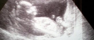
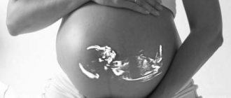
Ultrasound during pregnancy
The restructuring of the body functions of a pregnant woman is regulated by the central nervous system with the participation of the endocrine glands.
A woman’s reproductive organs undergo especially significant transformations during pregnancy:
- The vagina lengthens and expands: the folding of the mucous membrane increases, the tissues swell and acquire elasticity, which contributes to their better stretching during childbirth.
- During gestation, the uterus not only increases in size - its shape, location, and consistency change. If at the beginning of pregnancy the weight of the uterus is about 50 g, then by the time of birth it gains weight up to 1200 g. The blood supply to the organ increases significantly, and the number of muscle fibers in the uterine wall increases.
- In the ovaries, the process of follicle maturation is inhibited, and ovulation does not occur.
- In the mammary glands, the volume of glandular tissue increases, and excretory ducts develop. The breasts begin to actively grow, the areolas and nipples acquire dark pigmentation and become more sensitive. Towards the end of pregnancy, colostrum may be released from the nipples.
The changes also affect other systems and organs of a woman carrying a child.
The increase in body weight of a pregnant woman is associated both with the growth of the fetus and with ongoing changes in the body of the woman herself. By the end of gestation, the average weight gain is 13 kg (variations from 8 to 18 kg are possible).
Due to the stimulation of the vagus nerve, which innervates many internal organs, a pregnant woman often experiences changes in taste and smell, nausea, vomiting, excessive salivation, and dizziness.
The increased load on the cardiovascular system is due to several factors:
- The heart takes a more horizontal position in the chest.
- Formation of the uteroplacental circulation.
- Increase in circulating blood mass.
- Limitation of diaphragm mobility.
- Increased intra-abdominal pressure.
- Compression of the inferior vena cava by the growing uterus.
During normal pregnancy, blood pressure decreases in the second trimester by 5-15 mmHg. column and returns to the original numbers before childbirth. When the pregnant uterus compresses the inferior vena cava, the venous outflow worsens, the cardiac output decreases, which can provoke collapse.
Changes in the central nervous system lead to a certain transformation of the behavioral reactions of a pregnant woman. This is manifested by emotional lability (tearfulness, irritability), increased fatigue, and daytime sleepiness.
To fully assimilate food, the expectant mother’s body produces more digestive enzymes. The concentration of proteins, fatty acids, and glucose increases in the blood. These nutrients, entering the baby’s blood through the placenta, provide the developing body with everything necessary for growth and development.
Pregnancy after IVF >>>
In the lungs, the level of oxygen saturation in the blood increases due to an increase in the number of red blood cells.
The urinary system is activated, which has to remove waste products from both the woman and the developing fetus. The tone of the bladder relaxes, which increases the frequency of the urge to urinate. Due to stagnation of urine, complications such as pyelonephritis and hydronephrosis can develop.
In later stages, the mobility of the pelvic joints increases, and the ability of the ligamentous apparatus to stretch increases. If the pelvic joints soften excessively, then divergence of the pubic bones may occur, which is manifested by pain and the appearance of the so-called “duck” gait.
Areas of hyperpigmentation appear on the skin: on the nipples, along the midline of the abdomen, on the navel, chloasma on the face. The functions of the sweat and sebaceous glands are enhanced. Sometimes there is increased hair growth, formation on the skin (chest, abdomen, thighs) - stretch marks - pregnancy scars, caused by the divergence of the deep layers of the skin.
Gastrulation process
After the appearance of the blastula, a new embryonic period begins in most multicellular organisms. It consists in the formation of the gastrula, which is represented by the following stages:
- Maturation of a two-layer embryo with the presence of endoderm and ectoderm.
- The formation of a three-layer embryo, in which the mesoderm grows in the form of a special leaf.
The indicated process is carried out through intussusception, with cells from the blastula from the marginal pole invaginating into the inner part. The outer covering layer is called ectoderm, the inner one is called endoderm. Mesoderm arises between them.
Complications of pregnancy
According to statistics, only 30-50% of pregnancies proceed physiologically, and this indicator shows a downward trend.
The most common pathological conditions complicating the course of pregnancy include:
- Miscarriages 15-20%.
- Premature birth 6-10%.
- Oligohydramnios, polyhydramnios – 8%.
- Late gestosis (preeclampsia, eclampsia) – 3-8%.
Complicated pregnancy can be caused by many factors on the part of the mother and the fetus.
Maternal complications
Somatic pathology. Pregnancy usually leads to decompensation of any chronic disease. The risk of developing late gestosis increases if a pregnant woman has hypertension, heart defects, glomerulonephritis, or pyelonephritis.
Endocrine pathology. With disorders of the hypothalamic-pituitary regulation and ovarian function, embryo implantation is disrupted and the contractile activity of the myometrium changes. All this threatens frozen pregnancy and early miscarriages.
Inflammatory diseases of the genitals increase the likelihood of developing an ectopic pregnancy, and chronic cervicitis provokes isthmic-cervical insufficiency and intrauterine infection of the fetus.
A history of pathological pregnancies and childbirth is a real threat of recurrent miscarriage.
Multiple pregnancy is also an increased risk factor for the development of complicated gestation. With it, a woman often worsens chronic somatic diseases, late toxicosis manifests itself, polyhydramnios develops, and there is a threat of premature termination of pregnancy.
Bacterial and viral infections suffered by a woman during pregnancy can cause birth defects, cause miscarriage or premature birth, and lead to intrauterine infection.
Pregnancies with genetic abnormalities incompatible with the life of the embryo result in early miscarriages. This triggers the mechanism of natural selection programmed by nature.
Immunological factors. Habitual miscarriage often occurs in women when the blood of the mother and baby is incompatible according to blood type or Rh factor.
Fetal complications
Pathology of embryonic structures during pregnancy: the umbilical cord, placenta, membranes of the fetal sac can lead to disruption of nutrition, gas exchange and the mechanisms of protection of the embryo and the development of fetoplacental insufficiency. Consequences: hypoxia, malnutrition, infection or congenital anomalies.
Rh conflict is characterized by the destruction of red blood cells with the development of severe complications, ranging from hemolytic jaundice requiring blood transfusion to fetal death.
Frozen pregnancy. If intrauterine death of the fetus occurs while it remains in the uterine cavity, there is a threat of the development of purulent-inflammatory processes: endometritis, peritonitis, sepsis, which without urgent help can lead to death.
Symptoms of a complicated pregnancy
The most common symptom of a complicated pregnancy is abdominal pain. It can be of a different nature: local or diffuse with irradiation to the groin and lower back. Pain syndrome is typical during ectopic pregnancy, threat of miscarriage or premature birth, or when there is a danger of uterine rupture (if there is a cesarean section scar on the uterus). In such cases, abdominal pain is accompanied by a deterioration in the general condition: weakness, dizziness, sometimes a drop in blood pressure, fainting.
Complications during pregnancy may also be indicated by discharge from the genital tract. Miscarriages, premature birth, placental abruption occur with bleeding of varying severity: from spotting to massive bleeding. Mucopurulent vaginal discharge is present during inflammatory processes. If the discharge is watery, it is most likely due to leakage or premature breaking of water.
In principle, any deterioration in the well-being of a pregnant woman can be regarded as a sign of a possible complication.
So, in the 1st trimester, symptoms such as weakness, nausea, vomiting are signs of early toxicosis.
Late gestosis causes dizziness, flashing “spots” before the eyes, swelling, nausea, vomiting, and a sharp rise in blood pressure.
Fever accompanies many infectious diseases and inflammatory processes.
With an exacerbation of chronic extragenital pathology, symptoms characteristic of this disease will come to the fore.
Complications of pregnancy from the fetus are identified by the nature of movement. With hypoxia, the fetus begins to actively move, and the absence of movement for 4 hours is a reason to immediately contact an obstetrician to find out the causes of this condition.
In order to prevent complications during pregnancy, a woman, even at the stage of pregnancy planning, must:
- undergo a full examination;
- make sure there are no contraindications to conception;
- undergo treatment of identified pathologies;
- to refuse from bad habits;
- lead a healthy lifestyle;
- register with the antenatal clinic on time;
- follow all recommendations of the obstetrician.
Known Factors
There are a number of exposures that can critically affect fetal development. Conventionally, they are divided into factors coming from the environment, and the lifestyle of a pregnant woman, past diseases. The first group includes the following manifestations:
- Radiation emission. If it is damaged at the beginning of the embryonic period, in most cases spontaneous miscarriage occurs.
- Poisoning with chemical components, such as dyes, fertilizers, benzene, and chemotherapy.
- Electromagnetic radiation that affects you when you are in close proximity to switched on electrical appliances.
Embryonic development is often disrupted directly by a pregnant woman who has suffered genetic or chromosomal diseases, infectious lesions (herpes, syphilis, rubella). The cause is alcohol and drug abuse at any stage. Among the dangerous diseases are obesity, bronchial asthma, and heart failure, due to which the normal supply of oxygen to the tissues of the embryo is disrupted.
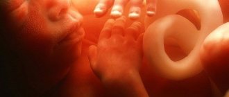
Embryo 5 - 6 weeks
Embryo. 5 weeks from the moment of fertilization.
At 5 weeks, the umbilical cord is formed, which will connect the baby to the placenta. The umbilical cord contains arteries and veins. The length of the umbilical cord by the end of childbirth can reach 70 cm. At the 5th week, the arms and legs look like flippers, but individual details can already be distinguished. The first impulses begin to be generated in the nervous system - the basis of nervous activity. The head of the embryo is formed, and holes for the ears, eyes, mouth and nose appear in it. The length of the embryo reaches 1 cm.
Embryo 8 weeks
The kidneys begin to produce urine. Hair follicles form in the skin. Almost all vital organs have already been formed. Reflexes and feelings are triggered. Ears form, the face no longer looks like an alien from a science fiction movie. It is clearly visible that a person is growing. The bones are still made of cartilage, but later they will be saturated with calcium and turn into real bone tissue. This process will be completely completed only by the age of 25, long after birth. The size of the embryo is almost 2 cm.
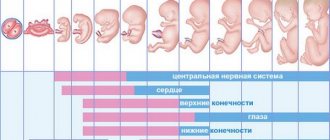
Dangerous periods
Having considered what processes occur at the stage of embryonic development, it is worth highlighting the most vulnerable and dangerous periods. Each of them is responsible for the formation of certain areas:
- at 2–11 weeks the brain is formed;
- at 3-7 weeks the heart and organs of vision begin to develop;
- at 3-8 weeks the limbs are identified;
- at week 9 the belly appears;
- At 4–12 weeks, the genital organs are formed.
The given characteristics of the complex embryonic period show how important all its stages are. The formation of tissues and organs of the future organism occurs in humans, animals (from mice to crocodile), and all multicellular creatures. Embryogenesis lasts in different ways, which should be briefly indicated in a message on biology, and the information should be collected in a table.
Embryo 11 weeks
The baby has a hairstyle. The fine hair on the head and body is called lanugo. They usually fall out by the time of birth. The skin is almost transparent, blood vessels are visible. The first foci of ossification form in the bones. White blood cells - leukocytes - appear in the blood. The arms are pulled up to the face, the baby can put a finger in the mouth. We begin to do exercises - the baby can actively move in the womb, but its size (10-15 cm) and low weight (30 g) do not yet allow the mother to properly feel these movements.
Embryo. 6 weeks from the moment of fertilization.
At week 6, it is already possible to distinguish the features of the future face. Limbs and fingers are developing. The baby begins to make his first movements. The pigment that determines the color of the eyes is formed on the iris. At week 6, the embryo's heartbeat can be seen and heard using an ultrasound scanner or ultrasound. The placenta is fully formed. The rudiments of the lungs, kidneys, gonads, stomach and intestines are formed. Amniotic fluid already surrounds the embryo. The 6th week of pregnancy in Western countries is considered the optimal time for the first visit to a gynecologist. It is necessary to do an ultrasound, undergo a series of tests, and undergo an examination by specialized specialists.
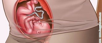
Embryo 20 weeks -30 weeks
Did you feel the kick? Now the baby communicates with his mother this way. If mom is sad, upset about something, or there is loud music around, it’s smoky, stuffy, the baby violently expresses his opinion. At first it’s timid, but by the end of pregnancy this method of communication will no longer cause tenderness. Constantly getting kicked from the inside can be quite painful. A sudden noise or fright of the mother leads to a similar response from the child. Protective reflexes arise.
In the lungs, the formation of respiratory alveoli is completed. Fingerprints form on hands. Brain development occurs at the fastest rate. Neural connections are formed. The baby can blink. The immune system is formed. The respiratory organs are developed enough to provide breathing in the event of premature birth, but sometimes only with medical assistance and special equipment. Adipose tissue appears. By 30 weeks, growth reaches 40-43 cm, weight up to 1.5 kg.