The human body works as a single system, where each organ performs its own function. The heart is the main organ of the circulatory system and is responsible for filling all the blood vessels in the body.
If the structure of the heart chambers or large vessels differs from normal, this indicates a defect. But how to determine the presence of a heart defect - congenital or acquired (CHD and PPS)? Is it possible to understand by feeling that it’s time to see a cardiologist? See symptoms of congenital heart disease in newborns? What procedures will help doctors recognize the disease and based on what signs? We will tell you all about the manifestations and symptoms of heart defects in adults and children, possible patient complaints and modern diagnostic methods!
Etiology
Congenital heart defect
Heart defects in newborns can develop due to:
- various mutations at the gene level;
- unfavorable environmental situation in the area where the pregnant woman lives;
- the woman has a history of abortions, miscarriages, and stillborn children;
- the use of certain groups of pharmaceuticals during pregnancy. Antibiotics, antiviral and other drugs with strong effects pose a particular danger to the fetus;
- hereditary predisposition. The risk that a baby will develop a heart defect increases many times if the pregnant woman has close relatives with the same pathology;
- illnesses of an infectious nature that a woman suffered while carrying a child. Particularly dangerous include cytomegaly, rubella, and herpes. The risk especially increases if these pathologies affect a woman in the early stages of pregnancy. The fact is that it is during this period that all organs are formed;
- age of the pregnant woman. Scientists have noticed a trend that the older a woman is, the higher the likelihood that she will have a child with a heart defect. Currently, the risk group includes representatives of the fair sex who have crossed the 35-year mark;
- strong x-ray training;
- consumption of large doses of alcoholic beverages by a woman while carrying a child. Recently, this reason has come to the fore in the development of heart defects. Alcohol has a detrimental effect not only on the mother’s body, but also on the body of her unborn baby.
Possible cures
Radical treatment of heart defects found in newborns consists only of surgical intervention, since drug therapy only makes it possible to relieve the intensity of symptoms and temporarily improve the quality of life of a little person.
Typically, medications are used to prepare the child's body for subsequent surgical violence.
Open operations are performed if treatment of severe defects of a combined type is required, when a huge amount of surgical intervention must be performed at once.
Such procedures are prescribed:
- patients whose health condition allows them to wait their turn for surgery, which can last a year or more;
- children whose defect needs to be corrected in the next six months;
- patients requiring treatment for a maximum of a couple of weeks;
- children with severe congenital heart disease who may die in the next 12-24 hours.
It is unfortunate, but there are children who from birth have only one ventricle, no heart septum, or underdeveloped valves. These babies can only count on palliative intervention, in which their health status improves temporarily.
In cases where minor heart defects are observed, doctors recommend that parents follow a wait-and-see approach. For example, a completely open ductus arteriosus is quite capable of closing on its own a few months after the child is born.
And such an anomaly as an aortic valve with two leaflets does not show its presence at all, which completely negates the need for drug or surgical intervention.
Symptoms
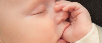
Symptoms indicating a heart defect in newborns directly depend on the type of defect, as well as on the severity of the pathological process. Small-sized defects practically do not manifest themselves at all, which greatly complicates their timely diagnosis. But it is worth noting that even severe forms of anomalies can be completely asymptomatic, which often causes the death of a newborn in the first days of his life. It is possible to save the life of a baby with severe defects that prevent the heart from functioning normally only through surgical intervention. Conservative therapy is out of the question.
The main signs that may indicate the presence of abnormalities in the baby’s cardiovascular system:
- increased respiratory movements per minute;
- the formation of edema (especially in the legs);
- weakness;
- lethargy;
- the child sucks weakly at the breast and may even refuse it completely;
- severe tachycardia;
- frequent regurgitation;
- cyanosis. It is especially pronounced on the limbs and in the area of the nasolabial triangle;
- heart murmurs. They can only be identified by a qualified doctor during auscultation.
Clinical picture of the development of PPS
PPS is otherwise called valve defects: in these diseases, it is the heart valves that are affected. The reasons for their development are infections, inflammation, autoimmune processes, overload of the heart chambers.
Let us briefly consider the classification of these diseases.
By localization:
- Monovalve - only one valve is affected.
- Combined - more than one valve is affected: two-valve, three-valve.
According to the functional form:
- Simple – stenosis or insufficiency.
- Combined - combine several simple defects on several valves.
- Combined - stenosis and insufficiency of only one of the valves.
Due to the development (etiology), diseases are rheumatic (up to 30-50% of all mitral stenoses are consequences of rheumatism), atherosclerotic, caused by bacterial endocarditis, syphilis (syphilitic heart disease is included in the list of rare cardiopathologies) and other diseases.
If the defects are minimally expressed, they do not appear clinically. In the stages of decompensation, hemodynamic disturbances appear, which are characterized by shortness of breath during physical activity, bluish skin, swelling, tachycardia, cough, and chest pain.
Let's take a closer look at the symptoms of acquired heart defects: how do they manifest themselves?
Mitral valve insufficiency and stenosis
In the stage of compensation for mitral regurgitation, people do not feel problems , but as the condition worsens, shortness of breath (first during physical activity, then at rest), palpitations, dry cough, and chest pain (in the area of the heart) may occur. Later, swelling of the lower extremities and pain in the right hypochondrium appear.
During examination, doctors reveal cyanosis of the skin and swelling of the veins in the neck. When listening, a weakening or absence of the first tone and systolic murmur are noticed. There are no characteristic changes in pulse and blood pressure.
With mitral stenosis, new complaints are added to the above . A person who suddenly stands up may develop cardiac asthma. The cough is dry, there may be some sputum, and hemoptysis occurs. The voice becomes hoarse, and increased fatigue occurs. Often, against the background of heart pain and tachycardia, arrhythmia begins - interruptions in rhythm.
What will the doctor see? On pale skin, a sharply defined bluish “blush” appears - a triangle from the tip of the nose to the lips. During auscultation, you can hear the so-called three-part “quail rhythm”, protodiastolic and presystolic murmur. Hypotension is possible (pressure tends to decrease), pulse varies depending on the location of measurement.
Aortic stenosis and insufficiency
Aortic stenosis occurs without symptoms for a long time; the first complaints begin when the valve opening narrows to more than 2/3 of the normal state . These are pains of a compressive nature in the chest during physical activity, fainting, dizziness.
Later, cardiac asthma, shortness of breath at rest, fatigue and weakness may develop. Further development causes swelling of the legs and pain in the hypochondrium on the right.
The doctor will also see external signs of the defect: pale or blue discoloration of the skin, swelling of the neck veins. Pay attention to systolic trembling a la a cat's purr, weakening of the first and second tones, systolic murmur, which increases in a lying position on the right side if you hold your breath while exhaling.
Pulse rare, weak. Systolic pressure is low, diastolic pressure is normal or increased.
With aortic insufficiency, there are practically no complaints during compensation; sometimes tachycardia and pulsation behind the sternum are observed . In the stage of decompensation, angina pain in the chest occurs, for which nitroglycerin does not help well, and standard symptoms: dizziness, fainting, shortness of breath (first with exertion, then at rest), swelling, a feeling of heaviness or pain on the right under the ribs.
The examination reveals pallor, pulsation of the peripheral arteries, rhythmic changes in skin color under the nails and on the lips with light pressure, and possible head shaking synchronous with the pulse. On auscultation, organic and functional noises will be heard; listening to the femoral artery will show a double Traube tone and a double Vinogradov-Durozier murmur.
The pulse is accelerated and high. Systolic and pulse pressure increase, diastolic decreases.
Degrees
The degree of the disease is determined depending on the severity of the symptoms. In total, clinicians distinguish 4 of them:
Grade 1 – the baby’s condition is relatively stable. Cardiac activity is within normal limits. Typically, no specific treatment is required at this stage;
Stage 2 – symptoms gradually increase. Problems arise with feeding the child, and respiratory function is also impaired;
3rd degree - the clinic is supplemented by neurological manifestations, since the brain is not sufficiently supplied with blood;
4th degree – terminal. If it progresses, the patient experiences depression of respiratory and cardiac activity. It usually ends in death.
Congenital heart defects (CHD) in children
The formation of the fetal heart occurs towards the end of the first trimester of pregnancy. Using a method such as ultrasound, most congenital heart defects can be detected already at 16-18 weeks. The diagnosis is made definitively in the 2nd or 3rd trimester. With congenital heart disease, children may have cyanosis if the amount of reduced erythrocyte hemoglobin is increased to 50 g/l. This is influenced by factors such as:
- an increase in the amount of venous blood entering the systemic circulation in the presence of blood discharge from the right parts of the heart to the left
- degree of blood oxygenation in the lungs
- degree of oxygen utilization by tissues
Cyanosis affects changes in peripheral blood: polycythemia and hyperhemoglobinemia. Chronic oxygen deficiency causes changes in the nail phalanges - the fingers resemble drumsticks.
There are 3 phases of the course of congenital heart defects :
The first phase is when reactions of adaptation and compensation to disturbances in the dynamics of blood circulation occur. If hemodynamics are significantly impaired, unstable myocardial hyperfunction appears, an emergency option according to V.V. Larin and F. Z. Meerson, so decompensation easily develops.
The second phase is relative compensation. The child’s physical development improves, as does his motor activity.
The third phase is terminal. It occurs when compensatory capabilities are exhausted and dystrophic and degenerative changes develop in the heart muscle and parenchymal organs. Various diseases and complications bring the development of this phase of the disease closer, which necessarily ends in death.
F. 3. Meerson and his colleagues distinguish 3 stages of compensatory hyperfunction of the heart :
1. Emergency
The intensity of functioning of myocardial structures increases. Signs of acute heart failure appear. The exchange change appears.
2. Second stage
The normal intensity of functioning of the myocardial structures is recorded. Disorders of metabolism, structure and regulation of the heart progress.
3. Stage of progressive cardiosclerosis and gradual exhaustion
The intensity of synthesis of nucleic acids and proteins in the hypertrophied myocardium decreases.
During the period of relative compensation, the syndrome of capillary-trophic insufficiency of the hemomicrocirculation system gradually develops, leading to a discrepancy between transcapillary blood flow and cardiac output. As a consequence of this, metabolic disorders appear in tissues, as well as dystrophic, atrophic and sclerotic changes in internal organs.
Diagnostics
Today, the most informative method that makes it possible to identify the presence of anomalies in the structure of the heart is ECHO cardiography. This method gives the doctor the opportunity to assess the condition of all elements of the heart - chambers, septa, valves, holes. Doctors also often resort to Doppler ultrasound. The method makes it possible to obtain information about the intensity of blood flow and its turbulence.
Additional diagnostic methods:
- ECG;
- radiography;
- MRI of the heart;
- CT scan of the heart.
The process of clarifying and making a diagnosis
If a child is suspected of having a heart defect, he is immediately sent to see a cardiologist or immediately to a cardiac surgery center. Its employees once again check all the symptoms and identify them with all types of congenital heart disease, assess the nature of the pulse and blood pressure, study the condition of all systems and organs, and conduct appropriate studies and tests.
If the defect raises any doubts, the baby will have to undergo catheterization - insertion of a probe through the vessels.
Parents themselves are often indignant that the pathology was not established at the gestation stage, when it was possible to make a decision to terminate the pregnancy.
There may be several reasons for this situation:
- lack of professionalism of the antenatal clinic staff;
- imperfection of equipment used to conduct standard and planned studies;
- natural structural features of the heart and blood vessels of the fetus, which make it difficult to see the presence of an anomaly.